Department of Biomedical Imaging and Image-guided Therapy, Medical University of Vienna, Waehringer Guertel 18-20, 1090, Vienna, Austria.
Comprehensive Cancer Center, Medical University of Vienna, Waehringer Guertel 18-20, 1090, Vienna, Austria.
J Neurooncol. 2018 Oct;140(1):173-178. doi: 10.1007/s11060-018-2948-8. Epub 2018 Jul 14.
The purpose of this study was to evaluate the prognostic relevance of temporal muscle thickness (TMT) in melanoma patients with newly diagnosed brain metastases.
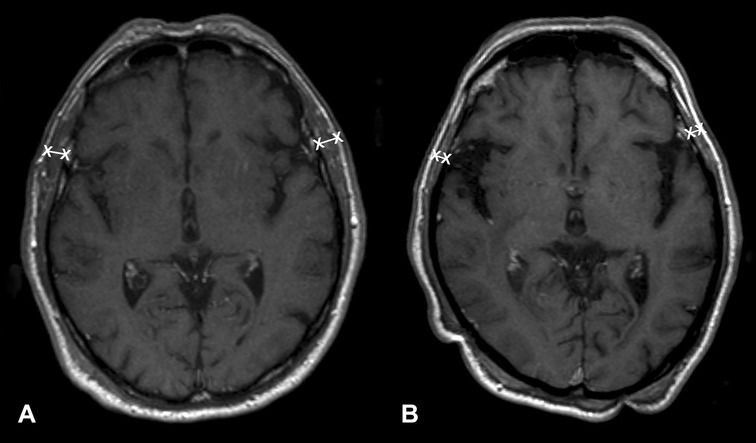
TMT was retrospectively assessed in 146 melanoma patients with newly diagnosed brain metastases on cranial magnetic resonance images. Chart review was used to retrieve clinical parameters, including disease-specific graded prognostic assessment (DS-GPA) and survival times.
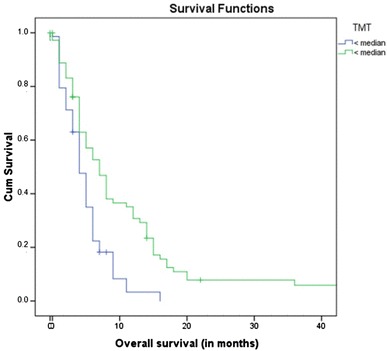
Patients with a TMT > median showed a statistically significant increase in survival time (13 months) compared to patients with a TMT < median (5 months; p < 0.001; log rank test). A Cox regression model revealed that the risk of death was increased by 27.9% with every millimeter reduction in TMT. In the multivariate analysis, TMT (HR 0.724; 95% 0.642-0.816; < 0.001) and DS-GPA (HR 1.214; 95% CI 1.023-1.439; p = 0.026) showed a statistically significant correlation with overall survival.
TMT is an independent predictor of survival in melanoma patients with brain metastases. This parameter may aid in patient selection for clinical trials or to the choice of different treatment options based on the determination of frail patient populations.
本研究旨在评估新诊断脑转移的黑色素瘤患者颞肌厚度(TMT)的预后相关性。
回顾性分析了 146 例新诊断脑转移的黑色素瘤患者的颅磁共振图像上的 TMT。通过图表回顾检索临床参数,包括疾病特异性预后评估分级(DS-GPA)和生存时间。
TMT 值大于中位数的患者的生存时间(13 个月)与 TMT 值小于中位数的患者(5 个月)相比,具有统计学显著差异(p<0.001;对数秩检验)。Cox 回归模型显示,TMT 每减少 1 毫米,死亡风险增加 27.9%。在多变量分析中,TMT(HR 0.724;95%CI 0.642-0.816;<0.001)和 DS-GPA(HR 1.214;95%CI 1.023-1.439;p=0.026)与总生存时间呈统计学显著相关。
TMT 是黑色素瘤脑转移患者生存的独立预测因子。该参数可能有助于患者选择临床试验或根据确定虚弱患者群体选择不同的治疗选择。