• 文献检索
  • 文档翻译
  • 深度研究
  • 学术资讯
  • Suppr Zotero 插件Zotero 插件
  • 邀请有礼
  • 套餐&价格
  • 历史记录
应用&插件
Suppr Zotero 插件Zotero 插件浏览器插件Mac 客户端Windows 客户端微信小程序
定价
高级版会员购买积分包购买API积分包
服务
文献检索文档翻译深度研究API 文档MCP 服务
关于我们
关于 Suppr公司介绍联系我们用户协议隐私条款
关注我们

Suppr 超能文献

核心技术专利:CN118964589B侵权必究
粤ICP备2023148730 号-1Suppr @ 2026

文献检索

告别复杂PubMed语法,用中文像聊天一样搜索,搜遍4000万医学文献。AI智能推荐,让科研检索更轻松。

立即免费搜索

文件翻译

保留排版,准确专业,支持PDF/Word/PPT等文件格式,支持 12+语言互译。

免费翻译文档

深度研究

AI帮你快速写综述,25分钟生成高质量综述,智能提取关键信息,辅助科研写作。

立即免费体验

评估中心静脉导管的“快速心房涡流征”:经过有限培训的住院医师的表现。

The "rapid atrial swirl sign" for assessing central venous catheters: Performance by medical residents after limited training.

机构信息

Department of Nephrology and Rheumatology, University Medical Center Goettingen, Goettingen, Germany.

Department of Gastroenterology and Gastrointestinal Oncology, University Medical Center Goettingen, Goettingen, Germany.

出版信息

PLoS One. 2018 Jul 16;13(7):e0199345. doi: 10.1371/journal.pone.0199345. eCollection 2018.

DOI:10.1371/journal.pone.0199345
PMID:30011285
原文链接:https://pmc.ncbi.nlm.nih.gov/articles/PMC6047781/
Abstract

RATIONALE

Central venous catheter (CVC) placement is a standard procedure in critical care. Ultrasound guidance during placement is recommended by current guidelines, but there is no consensus on the best method for evaluating the correct CVC tip position. Recently, the "rapid atrial swirl sign" (RASS) has been investigated in a limited number of studies.

OBJECTIVES

We performed a prospective diagnostic accuracy study of focused echocardiography for the evaluation of CVC tip position in our medical ICU and IMC units.

METHODS

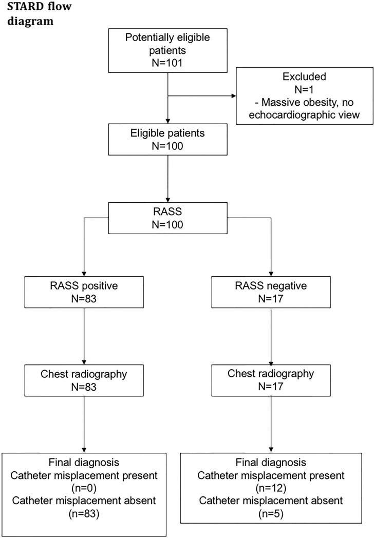
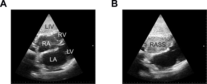
We performed a prospective diagnostic accuracy study in 100 patients admitted to the Intensive Care Unit and Intermediate Care Unit at our center. The first 10 subjects were assessed by one staff physician investigator (reference cohort), the remaining 90 patients by different residents (test cohort). All patients received a post-procedural chest radiograph (CXR) as gold standard. CVC placement was assessed with focused echocardiography performed by residents after a short training session. A rapid opacification of the right atrium (RASS) after injection of 10 mL of normal saline was regarded as "positive", flush after more than two seconds was defined as "delayed", no flush was a "negative" test result.

MEASUREMENTS AND MAIN RESULTS

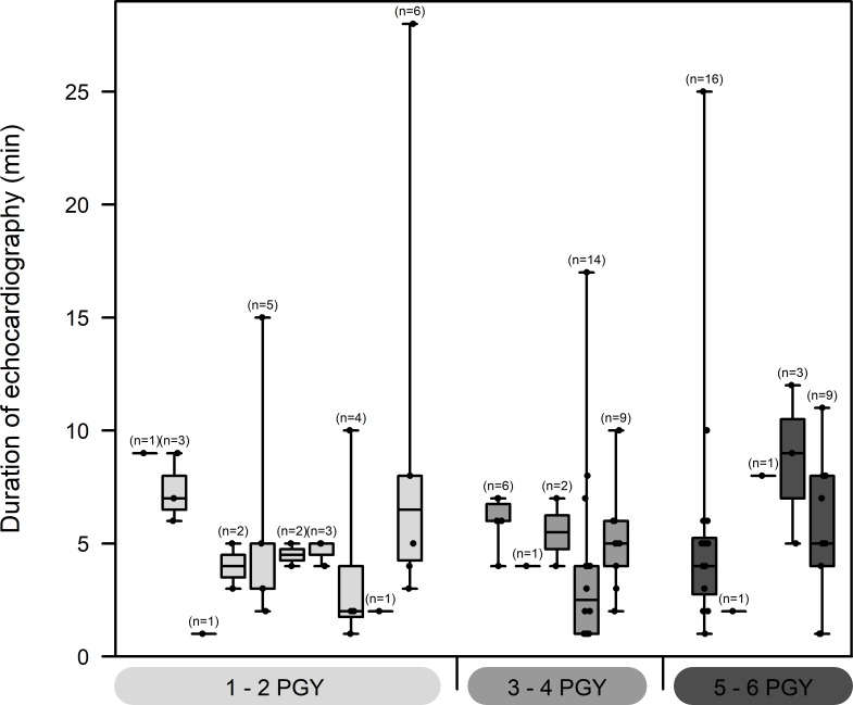
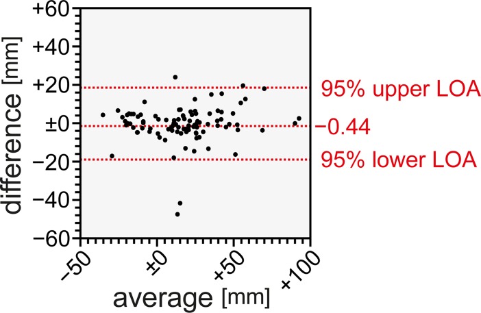
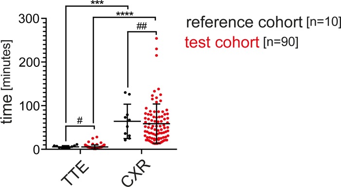
Overall sensitivity of the RASS was 100% (95% CI 73.54-100%), specificity was 94.32% (CI 87.24-98.13%). Positive and negative predictive values were 70.59% (CI 44.04-89.09%) and 100% (CI 95.65-100%), respectively. Median time for echocardiographic testing was 5 minutes (1-28) in the whole cohort, CXRs were available after 49.5 minutes (13-254). Interrater agreement of the RASS was 0.77 (Cohen's kappa), Measurement of CVC tip position was not different between two observers. Test characteristics were similar among differently experienced residents.

CONCLUSIONS

Presence of the RASS by focused echocardiography showed excellent sensitivity and specificity and was equally performed by residents after minimal training. In patients with a positive RASS, routine CXR can be safely omitted, reducing time, costs and radiation exposure. A negative RASS should lead to a search for misplaced catheters.

CLINICAL TRIAL REGISTRATION

The study was registered with www.clinicaltrials.gov (NCT02661607).

摘要

背景

中心静脉导管(CVC)置管术是重症监护中的一项标准操作。目前的指南建议在置管过程中进行超声引导,但对于评估正确的 CVC 尖端位置的最佳方法尚无共识。最近,在有限数量的研究中已经研究了“快速心房漩涡征”(RASS)。

目的

我们对我院重症监护病房(ICU)和中级监护病房(IMC)进行了一项经胸超声心动图评估 CVC 尖端位置的前瞻性诊断准确性研究。

方法

我们对我院中心收治的 100 例 ICU 和 IMC 患者进行了一项前瞻性诊断准确性研究。前 10 例由一名主治医生调查员(参考队列)评估,其余 90 例由不同的住院医师(测试队列)评估。所有患者均接受术后胸部 X 线摄影(CXR)作为金标准。在短时间的培训后,住院医师使用经胸超声心动图评估 CVC 置管。在注入 10ml 生理盐水后右心房快速显影(RASS)为“阳性”,超过两秒后显影为“延迟”,无显影为“阴性”。

测量和主要结果

总体而言,RASS 的敏感性为 100%(95%CI 73.54-100%),特异性为 94.32%(CI 87.24-98.13%)。阳性和阴性预测值分别为 70.59%(CI 44.04-89.09%)和 100%(CI 95.65-100%)。整个队列的超声心动图检查中位时间为 5 分钟(1-28 分钟),CXR 可在 49.5 分钟(13-254 分钟)后获得。RASS 的观察者间一致性为 0.77(Cohen's kappa),两名观察者测量 CVC 尖端位置无差异。不同经验的住院医师的测试特征相似。

结论

经胸超声心动图显示 RASS 阳性具有极好的敏感性和特异性,且在经过最少的培训后,住院医师也能同样出色地进行检查。在 RASS 阳性的患者中,常规 CXR 可以安全省略,从而减少时间、成本和辐射暴露。RASS 阴性应提示寻找位置不当的导管。

临床试验注册

该研究在 www.clinicaltrials.gov 上注册(NCT02661607)。

https://cdn.ncbi.nlm.nih.gov/pmc/blobs/2831/6047781/33215e60466d/pone.0199345.g006.jpg
https://cdn.ncbi.nlm.nih.gov/pmc/blobs/2831/6047781/6c80693c19e5/pone.0199345.g001.jpg
https://cdn.ncbi.nlm.nih.gov/pmc/blobs/2831/6047781/55221709cf41/pone.0199345.g002.jpg
https://cdn.ncbi.nlm.nih.gov/pmc/blobs/2831/6047781/53d05e503ec0/pone.0199345.g003.jpg
https://cdn.ncbi.nlm.nih.gov/pmc/blobs/2831/6047781/5da117a0db5c/pone.0199345.g004.jpg
https://cdn.ncbi.nlm.nih.gov/pmc/blobs/2831/6047781/088eee681b82/pone.0199345.g005.jpg
https://cdn.ncbi.nlm.nih.gov/pmc/blobs/2831/6047781/33215e60466d/pone.0199345.g006.jpg

相似文献

1
The "rapid atrial swirl sign" for assessing central venous catheters: Performance by medical residents after limited training.评估中心静脉导管的“快速心房涡流征”:经过有限培训的住院医师的表现。
PLoS One. 2018 Jul 16;13(7):e0199345. doi: 10.1371/journal.pone.0199345. eCollection 2018.
2
Central vascular catheter placement evaluation using saline flush and bedside echocardiography.使用生理盐水冲洗和床旁超声心动图评估中心血管导管置入情况。
Acad Emerg Med. 2014 Jan;21(1):65-72. doi: 10.1111/acem.12283.
3
Prospective comparison of ultrasound and CXR for confirmation of central vascular catheter placement.超声与胸部X线用于确认中心血管导管置入位置的前瞻性比较。
Emerg Med J. 2016 Mar;33(3):176-80. doi: 10.1136/emermed-2015-205000. Epub 2015 Oct 7.
4
Use of Bedside Ultrasonography and Saline Flush Technique for Evaluation of Central Venous Catheter Placement in Children.使用床旁超声检查和生理盐水冲洗技术评估儿童中心静脉导管置入情况
Artif Organs. 2018 Dec;42(12):1157-1163. doi: 10.1111/aor.13281. Epub 2018 Aug 22.
5
The bubble study: ultrasound confirmation of central venous catheter placement.气泡研究:中心静脉导管置入的超声确认
Am J Emerg Med. 2015 Mar;33(3):315-9. doi: 10.1016/j.ajem.2014.10.010. Epub 2014 Oct 13.
6
Is traditional reading of the bedside chest radiograph appropriate to detect intraatrial central venous catheter position?传统的床边胸部X线片阅片法是否适合用于检测心房内中心静脉导管的位置?
Chest. 2008 Sep;134(3):527-533. doi: 10.1378/chest.07-2687. Epub 2008 Jul 18.
7
"Rapid Atrial Swirl Sign": A Better Tool Than the Landmark Technique for Ensuring Correct Depth of Insertion of Central Venous Catheters.“快速心房漩涡征”:一种比标志性技术更有助于确保中心静脉导管正确插入深度的工具。
Cureus. 2024 Jul 23;16(7):e65211. doi: 10.7759/cureus.65211. eCollection 2024 Jul.
8
A Simple and Safe Method for Checking the Position of Central Venous Catheters-A New and Reliable Threshold for Right Atrial Swirl Sign in Microbubbles Tests.一种简单安全的中心静脉导管位置检查方法——微泡试验中右心房漩涡信号的新可靠阈值
J Clin Med. 2024 Mar 14;13(6):1657. doi: 10.3390/jcm13061657.
9
Using central venous pressure waveform to confirm the placement of an internal jugular central venous catheter in the intensive care unit.使用中心静脉压波形确认重症监护病房中颈内静脉中央静脉导管的位置。
Aust Crit Care. 2024 Jul;37(4):628-631. doi: 10.1016/j.aucc.2023.10.007. Epub 2023 Dec 7.
10
Confirmation of correct central venous catheter position in the preoperative setting by echocardiographic "bubble-test".术前通过超声心动图“气泡试验”确认中心静脉导管位置正确。
Minerva Anestesiol. 2015 Sep;81(9):989-1000. Epub 2014 Nov 6.

引用本文的文献

1
Ultrasound Versus Fluoroscopy for Catheter Tip Confirmation in Long-Term Vascular Access: A Prospective Observational Study.超声与荧光透视在长期血管通路中导管尖端确认的比较:一项前瞻性观察研究。
Cureus. 2025 Jun 23;17(6):e86604. doi: 10.7759/cureus.86604. eCollection 2025 Jun.
2
Transthoracic Echocardiography for Central Venous Catheter Tip Positioning in Children: An Observational Study Using Transesophageal Echocardiography as Reference Method.经胸超声心动图用于儿童中心静脉导管尖端定位:一项以经食管超声心动图为参考方法的观察性研究
Paediatr Anaesth. 2025 Apr;35(4):310-315. doi: 10.1111/pan.15059. Epub 2025 Jan 4.
3

本文引用的文献

1
EFSUMB Guidelines on Interventional Ultrasound (INVUS), Part VI - Ultrasound-Guided Vascular Interventions.欧洲超声医学与生物学联合会介入超声指南(INVUS),第六部分 - 超声引导下的血管介入
Ultraschall Med. 2016 Oct;37(5):473-476. doi: 10.1055/s-0035-1553450. Epub 2015 Oct 29.
2
Prospective comparison of ultrasound and CXR for confirmation of central vascular catheter placement.超声与胸部X线用于确认中心血管导管置入位置的前瞻性比较。
Emerg Med J. 2016 Mar;33(3):176-80. doi: 10.1136/emermed-2015-205000. Epub 2015 Oct 7.
3
Saline Flush Test: Can Bedside Sonography Replace Conventional Radiography for Confirmation of Above-the-Diaphragm Central Venous Catheter Placement?
"Rapid Atrial Swirl Sign": A Better Tool Than the Landmark Technique for Ensuring Correct Depth of Insertion of Central Venous Catheters.
“快速心房漩涡征”:一种比标志性技术更有助于确保中心静脉导管正确插入深度的工具。
Cureus. 2024 Jul 23;16(7):e65211. doi: 10.7759/cureus.65211. eCollection 2024 Jul.
4
Are routine chest radiographs still indicated after central line insertion? A scoping review.中心静脉置管后是否仍需进行常规胸部X线检查?一项范围综述。
J Intensive Care Soc. 2024 Feb 19;25(2):190-207. doi: 10.1177/17511437241227739. eCollection 2024 May.
5
A Simple and Safe Method for Checking the Position of Central Venous Catheters-A New and Reliable Threshold for Right Atrial Swirl Sign in Microbubbles Tests.一种简单安全的中心静脉导管位置检查方法——微泡试验中右心房漩涡信号的新可靠阈值
J Clin Med. 2024 Mar 14;13(6):1657. doi: 10.3390/jcm13061657.
6
Comparison between ultrasound and chest X-ray to confirm central venous catheter tip position.超声与胸部X线用于确认中心静脉导管尖端位置的比较。
SA J Radiol. 2023 Jun 29;27(1):2587. doi: 10.4102/sajr.v27i1.2587. eCollection 2023.
7
Point-of-Care Ultrasound Protocol for Insertion and Confirmation of Central Venous Catheter Placement.中心静脉导管置入与确认的床旁超声检查方案
Cureus. 2022 Sep 17;14(9):e29259. doi: 10.7759/cureus.29259. eCollection 2022 Sep.
8
Determinants of Tunneled Hemodialysis Catheter Implantation Time by Ultrasound Guidance: A Single-Center Cross-Sectional Study.超声引导下带隧道血液透析导管植入时间的决定因素:一项单中心横断面研究
J Clin Med. 2022 Jun 19;11(12):3526. doi: 10.3390/jcm11123526.
9
The rapid atrial swirl sign for ultrasound-guided tip positioning of antegrade-tunneled hemodialysis catheters: A cross-sectional pilot study.快速心房涡流征在超声引导下经皮隧道式血液透析导管尖端定位中的应用:一项横断面初步研究。
J Vasc Access. 2023 Nov;24(6):1407-1411. doi: 10.1177/11297298221088763. Epub 2022 Apr 20.
10
Retrospective Assessment of Patient and Catheter Characteristics Associated With Malpositioned Central Venous Catheters in Pediatric Patients.回顾性评估与儿科患者中心静脉导管位置不当相关的患者和导管特征。
Pediatr Crit Care Med. 2022 Mar 1;23(3):192-200. doi: 10.1097/PCC.0000000000002882.
生理盐水冲洗试验:床旁超声检查能否替代传统放射成像来确认膈上中心静脉导管的位置?
J Ultrasound Med. 2015 Jul;34(7):1295-9. doi: 10.7863/ultra.34.7.1295.
4
Ultrasound-Guided Subclavian Vein Catheterization: A Systematic Review and Meta-Analysis.超声引导下锁骨下静脉置管术:系统评价和荟萃分析。
Crit Care Med. 2015 Jul;43(7):1498-507. doi: 10.1097/CCM.0000000000000973.
5
Confirmation of endovenous placement of central catheter using the ultrasonographic "bubble test".使用超声“气泡试验”确认中心静脉导管的静脉内放置。
Indian J Crit Care Med. 2015 Jan;19(1):38-41. doi: 10.4103/0972-5229.148642.
6
The bubble study: ultrasound confirmation of central venous catheter placement.气泡研究:中心静脉导管置入的超声确认
Am J Emerg Med. 2015 Mar;33(3):315-9. doi: 10.1016/j.ajem.2014.10.010. Epub 2014 Oct 13.
7
Confirmation of correct central venous catheter position in the preoperative setting by echocardiographic "bubble-test".术前通过超声心动图“气泡试验”确认中心静脉导管位置正确。
Minerva Anestesiol. 2015 Sep;81(9):989-1000. Epub 2014 Nov 6.
8
Looking through the retrospectoscope: reducing bias in emergency medicine chart review studies.回顾过往:减少急诊医学病历回顾研究中的偏倚
Ann Emerg Med. 2014 Sep;64(3):292-8. doi: 10.1016/j.annemergmed.2014.03.025. Epub 2014 Apr 18.
9
Central vascular catheter placement evaluation using saline flush and bedside echocardiography.使用生理盐水冲洗和床旁超声心动图评估中心血管导管置入情况。
Acad Emerg Med. 2014 Jan;21(1):65-72. doi: 10.1111/acem.12283.
10
The FLUSH study--flush the line and ultrasound the heart: ultrasonographic confirmation of central femoral venous line placement.FLUSH 研究--冲洗导管并超声心动:中心股静脉置管位置的超声确认。
Ann Emerg Med. 2014 Jun;63(6):678-83. doi: 10.1016/j.annemergmed.2013.12.020. Epub 2014 Jan 15.