• 文献检索
  • 文档翻译
  • 深度研究
  • 学术资讯
  • Suppr Zotero 插件Zotero 插件
  • 邀请有礼
  • 套餐&价格
  • 历史记录
应用&插件
Suppr Zotero 插件Zotero 插件浏览器插件Mac 客户端Windows 客户端微信小程序
定价
高级版会员购买积分包购买API积分包
服务
文献检索文档翻译深度研究API 文档MCP 服务
关于我们
关于 Suppr公司介绍联系我们用户协议隐私条款
关注我们

Suppr 超能文献

核心技术专利:CN118964589B侵权必究
粤ICP备2023148730 号-1Suppr @ 2026

文献检索

告别复杂PubMed语法,用中文像聊天一样搜索,搜遍4000万医学文献。AI智能推荐,让科研检索更轻松。

立即免费搜索

文件翻译

保留排版,准确专业,支持PDF/Word/PPT等文件格式,支持 12+语言互译。

免费翻译文档

深度研究

AI帮你快速写综述,25分钟生成高质量综述,智能提取关键信息,辅助科研写作。

立即免费体验

利用 X 射线相衬断层成像术对人体冠状动脉进行细胞分辨率的 3D 虚拟组织学研究。

Cellular-resolution 3D virtual histology of human coronary arteries using x-ray phase tomography.

机构信息

Department of Applied Physics, KTH Royal Institute of Technology/Albanova, Stockholm, Sweden.

Karolinska Institutet, Division of Cardiovascular Medicine, Department of Clinical Sciences, Danderyd University Hospital, Stockholm, Sweden.

出版信息

Sci Rep. 2018 Jul 20;8(1):11014. doi: 10.1038/s41598-018-29344-3.

DOI:10.1038/s41598-018-29344-3
PMID:30030461
原文链接:https://pmc.ncbi.nlm.nih.gov/articles/PMC6054690/
Abstract

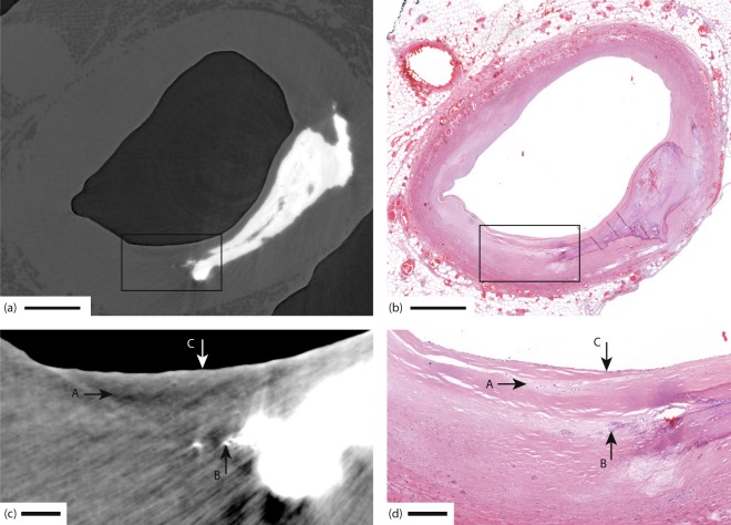
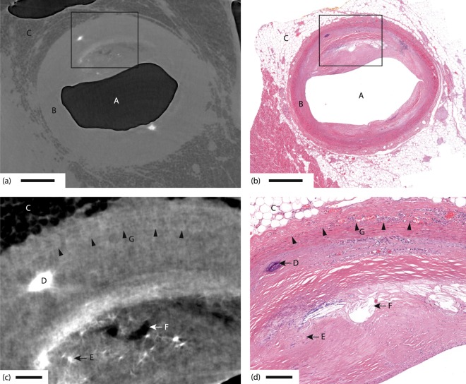
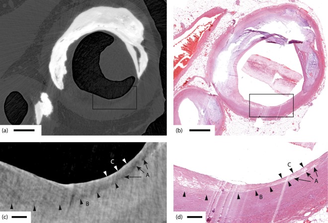
High-spatial-resolution histology of coronary artery autopsy samples play an important role for understanding heart disease such as myocardial infarction. Unfortunately, classical histology is often destructive, has thick slicing, requires extensive sample preparation, and is time-consuming. X-ray micro-CT provides fast nondestructive 3D imaging but absorption contrast is often insufficient, especially for observing soft-tissue features with high resolution. Here we show that propagation-based x-ray phase-contrast tomography has the resolution and contrast to image clinically relevant soft-tissue features in intact coronary artery autopsy samples with cellular resolution. We observe microscopic lipid-rich plaques, individual adipose cells, ensembles of few foam cells, and the thin fibrous cap. The method relies on a small-spot laboratory x-ray microfocus source, and provides high-spatial resolution in all three dimensions, fast data acquisition, minimum sample distortion and requires no sample preparation.

摘要

冠状动脉解剖样本的高空间分辨率组织学对于理解心肌梗死等心脏病具有重要作用。不幸的是,经典的组织学通常具有破坏性,切片较厚,需要广泛的样本制备,并且耗时。X 射线微计算机断层扫描(micro-CT)提供快速无损的 3D 成像,但吸收对比度通常不足,尤其是对于以高分辨率观察软组织特征。在这里,我们表明基于传播的 X 射线相衬层析成像具有分辨率和对比度,可以以细胞分辨率对完整冠状动脉解剖样本中的临床相关软组织特征进行成像。我们观察到微观富含脂质的斑块、单个脂肪细胞、少量泡沫细胞的集合体以及薄的纤维帽。该方法依赖于小光斑实验室 X 射线微焦点源,并在所有三个维度上提供高空间分辨率、快速数据采集、最小的样本变形,并且不需要样本制备。

https://cdn.ncbi.nlm.nih.gov/pmc/blobs/55ef/6054690/ff64d9a170ac/41598_2018_29344_Fig4_HTML.jpg
https://cdn.ncbi.nlm.nih.gov/pmc/blobs/55ef/6054690/468d89510610/41598_2018_29344_Fig1_HTML.jpg
https://cdn.ncbi.nlm.nih.gov/pmc/blobs/55ef/6054690/2174f60b1bb0/41598_2018_29344_Fig2_HTML.jpg
https://cdn.ncbi.nlm.nih.gov/pmc/blobs/55ef/6054690/ad467d35d8d8/41598_2018_29344_Fig3_HTML.jpg
https://cdn.ncbi.nlm.nih.gov/pmc/blobs/55ef/6054690/ff64d9a170ac/41598_2018_29344_Fig4_HTML.jpg

相似文献

1
Cellular-resolution 3D virtual histology of human coronary arteries using x-ray phase tomography.利用 X 射线相衬断层成像术对人体冠状动脉进行细胞分辨率的 3D 虚拟组织学研究。
Sci Rep. 2018 Jul 20;8(1):11014. doi: 10.1038/s41598-018-29344-3.
2
Integrating X-ray phase-contrast imaging and histology for comparative evaluation of breast tissue malignancies in virtual histology analysis.将 X 射线相衬成像与组织学相结合,用于虚拟组织学分析中对乳腺组织恶性肿瘤的对比评估。
Sci Rep. 2024 Mar 9;14(1):5831. doi: 10.1038/s41598-024-56341-6.
3
Three-dimensional virtual histology enabled through cytoplasm-specific X-ray stain for microscopic and nanoscopic computed tomography.通过针对细胞质的 X 射线染色实现微观和纳米级计算机断层扫描的三维虚拟组织学。
Proc Natl Acad Sci U S A. 2018 Mar 6;115(10):2293-2298. doi: 10.1073/pnas.1720862115. Epub 2018 Feb 20.
4
Three-dimensional imaging of whole mouse models: comparing nondestructive X-ray phase-contrast micro-CT with cryotome-based planar epi-illumination imaging.全鼠模型的三维成像:比较无损 X 射线相衬显微 CT 与基于冷冻切片的平面反射光照明成像。
J Microsc. 2014 Jan;253(1):24-30. doi: 10.1111/jmi.12094. Epub 2013 Nov 15.
5
Contrast enhancement for visualizing neuronal cytoarchitecture by propagation-based x-ray phase-contrast tomography.基于传播的 X 射线相位对比层析成像术对神经元细胞结构进行对比增强可视化。
Neuroimage. 2019 Oct 1;199:70-80. doi: 10.1016/j.neuroimage.2019.05.043. Epub 2019 May 23.
6
Extending two-dimensional histology into the third dimension through conventional micro computed tomography.通过传统的微型计算机断层扫描将二维组织学扩展到三维。
Neuroimage. 2016 Oct 1;139:26-36. doi: 10.1016/j.neuroimage.2016.06.005. Epub 2016 Jun 14.
7
Quantitative micro-CT based coronary artery profiling using interactive local thresholding and cylindrical coordinates.基于定量微型计算机断层扫描的冠状动脉轮廓分析:使用交互式局部阈值处理和柱面坐标
Technol Health Care. 2015;23(5):557-70. doi: 10.3233/THC-151010.
8
Propagation phase-contrast micro-computed tomography allows laboratory-based three-dimensional imaging of articular cartilage down to the cellular level.相衬增强的微观 CT 可以对关节软骨进行基于实验室的三维成像,达到细胞水平。
Osteoarthritis Cartilage. 2020 Jan;28(1):102-111. doi: 10.1016/j.joca.2019.10.007. Epub 2019 Oct 31.
9
High-Spatial-Resolution Three-dimensional Imaging of Human Spinal Cord and Column Anatomy with Postmortem X-ray Phase-Contrast Micro-CT.利用死后 X 射线相衬显微 CT 对人体脊髓和脊柱解剖结构进行高空间分辨率三维成像。
Radiology. 2021 Jan;298(1):135-146. doi: 10.1148/radiol.2020201622. Epub 2020 Oct 27.
10
3D vessel-wall virtual histology of whole-body perfused mice using a novel heavy element stain.使用新型重元素染色剂对全身灌注小鼠进行 3D 血管壁虚拟组织学研究。
Sci Rep. 2019 Jan 24;9(1):698. doi: 10.1038/s41598-018-36905-z.

引用本文的文献

1
Synchrotron-based X-ray 3D phase contrast imaging and analysis of transmural myocardial tissue from heart failure patients.基于同步加速器的X射线三维相衬成像及心力衰竭患者透壁心肌组织分析
Sci Rep. 2025 Jul 16;15(1):25867. doi: 10.1038/s41598-025-04012-5.
2
3D imaging of SARS-CoV-2 infected hamster lungs by X-ray phase contrast tomography enables drug testing.X 射线相位对比断层成像技术对感染 SARS-CoV-2 的仓鼠肺部进行 3D 成像,可用于药物测试。
Sci Rep. 2024 May 29;14(1):12348. doi: 10.1038/s41598-024-61746-4.
3
Phenotyping calcification in vascular tissues using artificial intelligence.

本文引用的文献

1
Early Detection and Treatment of the Vulnerable Coronary Plaque: Can We Prevent Acute Coronary Syndromes?易损冠状动脉斑块的早期检测与治疗:我们能否预防急性冠状动脉综合征?
Circ Cardiovasc Imaging. 2017 May;10(5). doi: 10.1161/CIRCIMAGING.116.005973.
2
Three-dimensional mouse brain cytoarchitecture revealed by laboratory-based x-ray phase-contrast tomography.基于实验室 X 射线相衬层析成像技术揭示的三维鼠脑细胞构筑
Sci Rep. 2017 Feb 27;7:42847. doi: 10.1038/srep42847.
3
High-resolution short-exposure small-animal laboratory x-ray phase-contrast tomography.
使用人工智能对血管组织中的钙化进行表型分析。
ArXiv. 2024 Jan 17:arXiv:2401.07825v2.
4
X-ray phase contrast reveals soft tissue and shell growth lines in mollusks.X 射线相衬成像揭示软体动物的软组织和壳生长线。
Commun Biol. 2024 Jan 3;7(1):17. doi: 10.1038/s42003-023-05457-y.
5
Acquisition of a single grid-based phase-contrast X-ray image using instantaneous frequency and noise filtering.使用瞬时频率和噪声滤波获取单个基于栅格的相衬 X 射线图像。
Biomed Eng Online. 2022 Dec 27;21(1):92. doi: 10.1186/s12938-022-01061-z.
6
Automated Machine-Learning Framework Integrating Histopathological and Radiological Information for Predicting IDH1 Mutation Status in Glioma.整合组织病理学和放射学信息以预测胶质瘤中异柠檬酸脱氢酶1(IDH1)突变状态的自动化机器学习框架
Front Bioinform. 2021 Oct 26;1:718697. doi: 10.3389/fbinf.2021.718697. eCollection 2021.
7
A groupwise registration and tractography framework for cardiac myofiber architecture description by diffusion MRI: An application to the ventricular junctions.基于扩散 MRI 的心肌纤维结构描述的分组配准和束追踪框架:在心室交界处的应用。
PLoS One. 2022 Jul 18;17(7):e0271279. doi: 10.1371/journal.pone.0271279. eCollection 2022.
8
Sub-micrometer morphology of human atherosclerotic plaque revealed by synchrotron radiation-based μCT-A comparison with histology.基于同步辐射的 μCT 揭示人动脉粥样硬化斑块的亚微米形态——与组织学的比较。
PLoS One. 2022 Apr 26;17(4):e0265598. doi: 10.1371/journal.pone.0265598. eCollection 2022.
9
Impact of intraventricular septal fiber orientation on cardiac electromechanical function.室间隔纤维方向对心机电功能的影响。
Am J Physiol Heart Circ Physiol. 2022 Jun 1;322(6):H936-H952. doi: 10.1152/ajpheart.00050.2022. Epub 2022 Mar 18.
10
Can laboratory x-ray virtual histology provide intraoperative 3D tumor resection margin assessment?实验室X线虚拟组织学能否提供术中三维肿瘤切除边缘评估?
J Med Imaging (Bellingham). 2022 May;9(3):031503. doi: 10.1117/1.JMI.9.3.031503. Epub 2022 Feb 7.
高分辨率短曝光小动物实验室 X 射线相衬层析成像。
Sci Rep. 2016 Dec 13;6:39074. doi: 10.1038/srep39074.
4
Correcting multi material artifacts from single material phase retrieved holo-tomograms with a simple 3D Fourier method.使用简单的三维傅里叶方法从单材料相检索的全息断层图中校正多材料伪影。
Opt Express. 2015 Dec 14;23(25):32718-27. doi: 10.1364/OE.23.032718.
5
AHA classification of coronary and carotid atherosclerotic plaques by grating-based phase-contrast computed tomography.基于光栅的相衬计算机断层扫描对冠状动脉和颈动脉粥样硬化斑块的美国心脏协会分类
Eur Radiol. 2016 Sep;26(9):3223-33. doi: 10.1007/s00330-015-4143-z. Epub 2015 Dec 17.
6
X-ray phase-contrast tomography for high-spatial-resolution zebrafish muscle imaging.用于高空间分辨率斑马鱼肌肉成像的X射线相衬断层扫描术
Sci Rep. 2015 Nov 13;5:16625. doi: 10.1038/srep16625.
7
X-ray phase-contrast computed tomography of human coronary arteries.X 射线相衬计算机断层扫描在冠状动脉成像中的应用。
Invest Radiol. 2015 Oct;50(10):686-94. doi: 10.1097/RLI.0000000000000169.
8
Complementary X-ray tomography techniques for histology-validated 3D imaging of soft and hard tissues using plaque-containing blood vessels as examples.使用含斑块的血管作为示例,用于对软组织和硬组织进行组织学验证的 3D 成像的补充 X 射线断层成像技术。
Nat Protoc. 2014;9(6):1401-15. doi: 10.1038/nprot.2014.091. Epub 2014 May 22.
9
Phase-contrast CT: qualitative and quantitative evaluation of atherosclerotic carotid artery plaque.相衬 CT:动脉粥样硬化颈动脉斑块的定性和定量评估。
Radiology. 2014 Jun;271(3):870-8. doi: 10.1148/radiol.14131554. Epub 2014 Mar 1.
10
Comparison of two x-ray phase-contrast imaging methods with a microfocus source.两种使用微焦点源的X射线相衬成像方法的比较。
Opt Express. 2013 Dec 16;21(25):30183-95. doi: 10.1364/OE.21.030183.