• 文献检索
  • 文档翻译
  • 深度研究
  • 学术资讯
  • Suppr Zotero 插件Zotero 插件
  • 邀请有礼
  • 套餐&价格
  • 历史记录
应用&插件
Suppr Zotero 插件Zotero 插件浏览器插件Mac 客户端Windows 客户端微信小程序
定价
高级版会员购买积分包购买API积分包
服务
文献检索文档翻译深度研究API 文档MCP 服务
关于我们
关于 Suppr公司介绍联系我们用户协议隐私条款
关注我们

Suppr 超能文献

核心技术专利:CN118964589B侵权必究
粤ICP备2023148730 号-1Suppr @ 2026

文献检索

告别复杂PubMed语法,用中文像聊天一样搜索,搜遍4000万医学文献。AI智能推荐,让科研检索更轻松。

立即免费搜索

文件翻译

保留排版,准确专业,支持PDF/Word/PPT等文件格式,支持 12+语言互译。

免费翻译文档

深度研究

AI帮你快速写综述,25分钟生成高质量综述,智能提取关键信息,辅助科研写作。

立即免费体验

使用新型重元素染色剂对全身灌注小鼠进行 3D 血管壁虚拟组织学研究。

3D vessel-wall virtual histology of whole-body perfused mice using a novel heavy element stain.

机构信息

Robarts Research Institute, The University of Western Ontario, London, Ontario, N6A 5B7, Canada.

Department of Medical Biophysics, The University of Western Ontario, London, Ontario, N6A 5C1, Canada.

出版信息

Sci Rep. 2019 Jan 24;9(1):698. doi: 10.1038/s41598-018-36905-z.

DOI:10.1038/s41598-018-36905-z
PMID:30679558
原文链接:https://pmc.ncbi.nlm.nih.gov/articles/PMC6345940/
Abstract

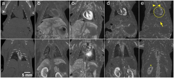
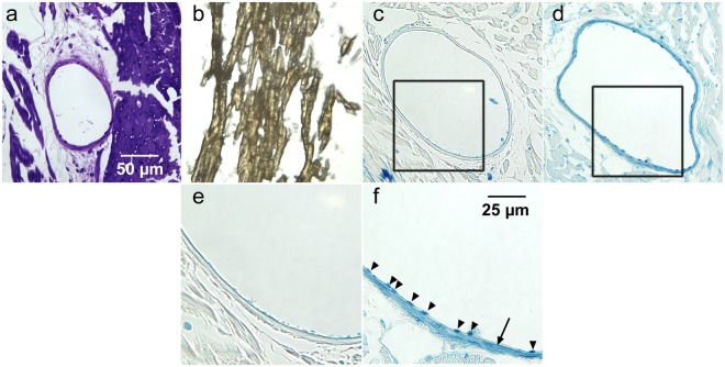
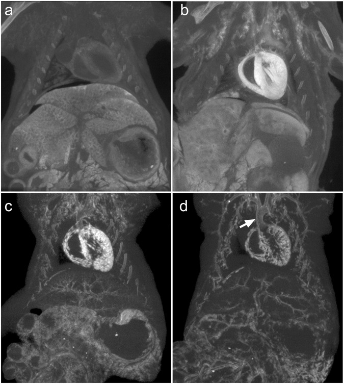
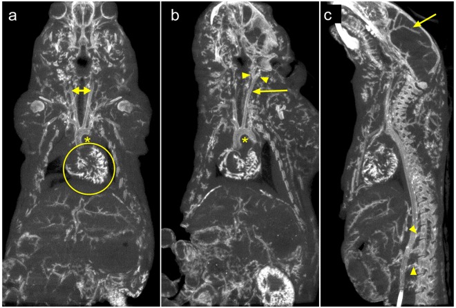
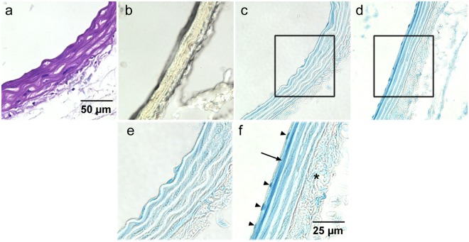
Virtual histology - utilizing high-resolution three-dimensional imaging - is becoming readily available. Micro-computed tomography (micro-CT) is widely available and is often coupled with x-ray attenuating histological stains that mark specific tissue components for 3D virtual histology. In this study we describe a new tri-element x-ray attenuating stain and perfusion protocol that provides micro-CT contrast of the entire vasculature of an intact mouse. The stain - derived from an established histology stain (Verhoeff's) - is modified to enable perfusion through the vasculature; the attenuating elements of the stain are iodine, aluminum, and iron. After a 30-minute perfusion through the vasculature (10-minute flushing with detergent-containing saline followed by 15-minute perfusion with the stain and a final 5-minute saline flush), animals are scanned using micro-CT. We demonstrate that the new staining protocol enables sharp delineation of the vessel walls in three dimensions over the whole body; corresponding histological analysis verified that the CT stain is localized primarily in the endothelial cells and media of large arteries and the endothelium of smaller vessels, such as the coronaries. The rapid perfusion and scanning protocol ensured that all tissues are available for further analysis via higher resolution CT of smaller sections or traditional histological sectioning.

摘要

虚拟组织学——利用高分辨率三维成像——正变得易于获得。微计算机断层扫描(micro-CT)广泛可用,并且通常与 X 射线衰减组织学染色结合使用,这些染色标记特定的组织成分以进行 3D 虚拟组织学。在这项研究中,我们描述了一种新的三元素 X 射线衰减染色和灌注方案,该方案可提供完整小鼠整个脉管系统的微 CT 对比。该染色剂——源自一种已建立的组织学染色剂(Verhoeff's)——经过改良后可使血管内的血液灌注;染色剂的衰减元素是碘、铝和铁。在血管内进行 30 分钟的灌注(用含洗涤剂的盐水冲洗 10 分钟,然后用染色剂灌注 15 分钟,最后用盐水冲洗 5 分钟)后,使用 micro-CT 对动物进行扫描。我们证明,新的染色方案能够在整个身体的三维空间中清晰地描绘血管壁;相应的组织学分析证实,CT 染色主要定位于大动脉的内皮细胞和中膜以及较小血管(如冠状动脉)的内皮细胞中。快速的灌注和扫描方案确保了所有组织都可通过较小切片的更高分辨率 CT 或传统的组织学切片进行进一步分析。

https://cdn.ncbi.nlm.nih.gov/pmc/blobs/ebf6/6345940/b063c04bc871/41598_2018_36905_Fig5_HTML.jpg
https://cdn.ncbi.nlm.nih.gov/pmc/blobs/ebf6/6345940/4bc32b724042/41598_2018_36905_Fig1_HTML.jpg
https://cdn.ncbi.nlm.nih.gov/pmc/blobs/ebf6/6345940/e3c58b4e1229/41598_2018_36905_Fig2_HTML.jpg
https://cdn.ncbi.nlm.nih.gov/pmc/blobs/ebf6/6345940/2539d53c7672/41598_2018_36905_Fig3_HTML.jpg
https://cdn.ncbi.nlm.nih.gov/pmc/blobs/ebf6/6345940/6871c0cdf014/41598_2018_36905_Fig4_HTML.jpg
https://cdn.ncbi.nlm.nih.gov/pmc/blobs/ebf6/6345940/b063c04bc871/41598_2018_36905_Fig5_HTML.jpg

相似文献

1
3D vessel-wall virtual histology of whole-body perfused mice using a novel heavy element stain.使用新型重元素染色剂对全身灌注小鼠进行 3D 血管壁虚拟组织学研究。
Sci Rep. 2019 Jan 24;9(1):698. doi: 10.1038/s41598-018-36905-z.
2
Three-dimensional imaging of the mouse heart and vasculature using micro-CT and whole-body perfusion of iodine or phosphotungstic acid.使用微型计算机断层扫描(micro-CT)对小鼠心脏和血管系统进行三维成像,并对碘或磷钨酸进行全身灌注。
Contrast Media Mol Imaging. 2014 Sep-Oct;9(5):383-90. doi: 10.1002/cmmi.1588. Epub 2014 Apr 25.
3
Retrograde perfusion and filling of mouse coronary vasculature as preparation for micro computed tomography imaging.小鼠冠状血管的逆行灌注和充盈,作为微型计算机断层扫描成像的准备。
J Vis Exp. 2012 Feb 10(60):e3740. doi: 10.3791/3740.
4
A perfusion procedure for imaging of the mouse cerebral vasculature by X-ray micro-CT.一种通过 X 射线微计算机断层扫描对小鼠脑血管进行成像的灌注方法。
J Neurosci Methods. 2014 Jan 15;221:70-7. doi: 10.1016/j.jneumeth.2013.09.002. Epub 2013 Sep 19.
5
Three-dimensional virtual histology enabled through cytoplasm-specific X-ray stain for microscopic and nanoscopic computed tomography.通过针对细胞质的 X 射线染色实现微观和纳米级计算机断层扫描的三维虚拟组织学。
Proc Natl Acad Sci U S A. 2018 Mar 6;115(10):2293-2298. doi: 10.1073/pnas.1720862115. Epub 2018 Feb 20.
6
Quantitative micro-CT based coronary artery profiling using interactive local thresholding and cylindrical coordinates.基于定量微型计算机断层扫描的冠状动脉轮廓分析:使用交互式局部阈值处理和柱面坐标
Technol Health Care. 2015;23(5):557-70. doi: 10.3233/THC-151010.
7
Coronary artery wall imaging in mice using osmium tetroxide and micro-computed tomography (micro-CT).使用锇四氧化物和微计算机断层扫描(micro-CT)对小鼠的冠状动脉壁成像。
J Anat. 2012 May;220(5):514-24. doi: 10.1111/j.1469-7580.2012.01483.x. Epub 2012 Feb 23.
8
Iodine-enhanced micro-computed tomography of atherosclerotic plaque morphology complements conventional histology.碘增强型显微计算机断层扫描对动脉粥样硬化斑块形态的研究补充了传统组织学方法。
Atherosclerosis. 2020 Nov;313:43-49. doi: 10.1016/j.atherosclerosis.2020.09.012. Epub 2020 Sep 24.
9
Cellular-resolution 3D virtual histology of human coronary arteries using x-ray phase tomography.利用 X 射线相衬断层成像术对人体冠状动脉进行细胞分辨率的 3D 虚拟组织学研究。
Sci Rep. 2018 Jul 20;8(1):11014. doi: 10.1038/s41598-018-29344-3.
10
3D Imaging and Quantitative Analysis of Vascular Networks: A Comparison of Ultramicroscopy and Micro-Computed Tomography.三维成像和血管网络定量分析:超微镜与微计算机断层扫描的比较。
Theranostics. 2018 Mar 7;8(8):2117-2133. doi: 10.7150/thno.22610. eCollection 2018.

引用本文的文献

1
Guidelines on antibody use in physiology research.抗体在生理学研究中的应用指南。
Am J Physiol Renal Physiol. 2024 Mar 1;326(3):F511-F533. doi: 10.1152/ajprenal.00347.2023. Epub 2024 Jan 18.
2
Regenerated Microvascular Networks in Ischemic Skeletal Muscle.缺血性骨骼肌中的再生微血管网络
Front Physiol. 2021 Jun 11;12:662073. doi: 10.3389/fphys.2021.662073. eCollection 2021.
3
A Review of Ex Vivo X-ray Microfocus Computed Tomography-Based Characterization of the Cardiovascular System.基于体外 X 射线微焦点计算机断层扫描的心血管系统特征评估综述。

本文引用的文献

1
X-ray based virtual histology allows guided sectioning of heavy ion stained murine lungs for histological analysis.基于 X 射线的虚拟组织学可用于引导对重离子染色的小鼠肺部进行切片,以进行组织学分析。
Sci Rep. 2018 May 16;8(1):7712. doi: 10.1038/s41598-018-26086-0.
2
Cathepsin B inhibition attenuates cardiovascular pathology in mucopolysaccharidosis I mice.组织蛋白酶B抑制作用可减轻黏多糖贮积症I型小鼠的心血管病变。
Life Sci. 2018 Mar 1;196:102-109. doi: 10.1016/j.lfs.2018.01.020. Epub 2018 Jan 31.
3
3D virtual histology of murine kidneys -high resolution visualization of pathological alterations by micro computed tomography.
Int J Mol Sci. 2021 Mar 23;22(6):3263. doi: 10.3390/ijms22063263.
4
X-ray Micro-Computed Tomography: An Emerging Technology to Analyze Vascular Calcification in Animal Models.X 射线微计算机断层扫描:一种分析动物模型血管钙化的新兴技术。
Int J Mol Sci. 2020 Jun 25;21(12):4538. doi: 10.3390/ijms21124538.
5
X-ray computed tomography in life sciences.X 射线计算机断层扫描在生命科学中的应用。
BMC Biol. 2020 Feb 27;18(1):21. doi: 10.1186/s12915-020-0753-2.
小鼠肾脏的 3D 虚拟组织学——通过微计算机断层扫描实现高分辨率的病理改变可视化。
Sci Rep. 2018 Jan 23;8(1):1407. doi: 10.1038/s41598-018-19773-5.
4
Dual-energy micro-CT for quantifying the time-course and staining characteristics of ex-vivo animal organs treated with iodine- and gadolinium-based contrast agents.双能 micro-CT 定量分析碘基和钆基对比剂处理的离体动物器官的时程和染色特征。
Sci Rep. 2017 Dec 12;7(1):17387. doi: 10.1038/s41598-017-17064-z.
5
Perivascular Adipose Tissue Angiotensin II Type 1 Receptor Promotes Vascular Inflammation and Aneurysm Formation.血管周脂肪组织血管紧张素 II 型 1 型受体促进血管炎症和动脉瘤形成。
Hypertension. 2017 Oct;70(4):780-789. doi: 10.1161/HYPERTENSIONAHA.117.09512. Epub 2017 Jul 31.
6
Effects of tissue fixation and dehydration on tendon collagen nanostructure.组织固定和脱水对肌腱胶原纳米结构的影响。
J Struct Biol. 2017 Sep;199(3):209-215. doi: 10.1016/j.jsb.2017.07.009. Epub 2017 Jul 28.
7
3D histopathological grading of osteochondral tissue using contrast-enhanced micro-computed tomography.使用对比增强微计算机断层扫描对骨软骨组织进行 3D 组织病理学分级。
Osteoarthritis Cartilage. 2017 Oct;25(10):1680-1689. doi: 10.1016/j.joca.2017.05.021. Epub 2017 Jun 9.
8
Differentiation of arterioles from venules in mouse histology images using machine learning.利用机器学习在小鼠组织学图像中区分小动脉和小静脉。
J Med Imaging (Bellingham). 2017 Apr;4(2):021104. doi: 10.1117/1.JMI.4.2.021104. Epub 2017 Feb 28.
9
Segmentation of digitized histological sections for quantification of the muscularized vasculature in the mouse hind limb.数字化组织学切片的分割,用于量化小鼠后肢的肌化血管系统。
J Microsc. 2017 Apr;266(1):89-103. doi: 10.1111/jmi.12522. Epub 2017 Feb 20.
10
μCT of ex-vivo stained mouse hearts and embryos enables a precise match between 3D virtual histology, classical histology and immunochemistry.对离体染色的小鼠心脏和胚胎进行微计算机断层扫描(μCT),可实现三维虚拟组织学、经典组织学和免疫化学之间的精确匹配。
PLoS One. 2017 Feb 8;12(2):e0170597. doi: 10.1371/journal.pone.0170597. eCollection 2017.