Schroeder Melanie M, Harrison Krystal R, Jaeckel Elizabeth R, Berger Hunter N, Zhao Xiwu, Flannery Michael P, St Pierre Emma C, Pateqi Nancy, Jachimska Agnieszka, Chervenak Andrew P, Wong Kwoon Y
Department of Ophthalmology & Visual Sciences, University of Michigan, Ann Arbor, MI, United States.
Department of Molecular, Cellular & Developmental Biology, University of Michigan, Ann Arbor, MI, United States.
Front Cell Neurosci. 2018 Jul 12;12:203. doi: 10.3389/fncel.2018.00203. eCollection 2018.
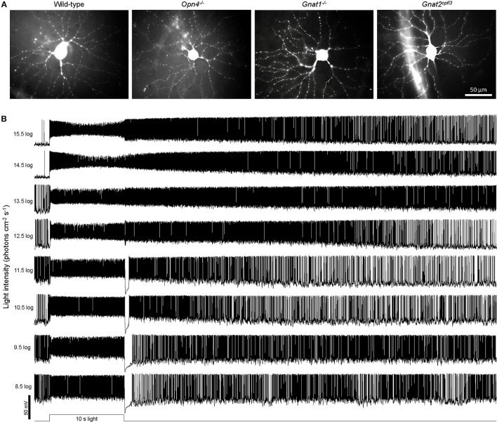
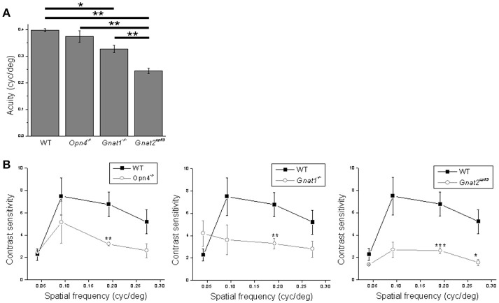
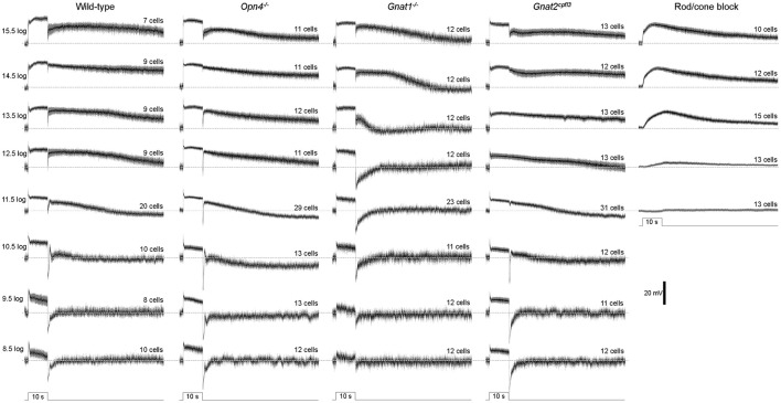
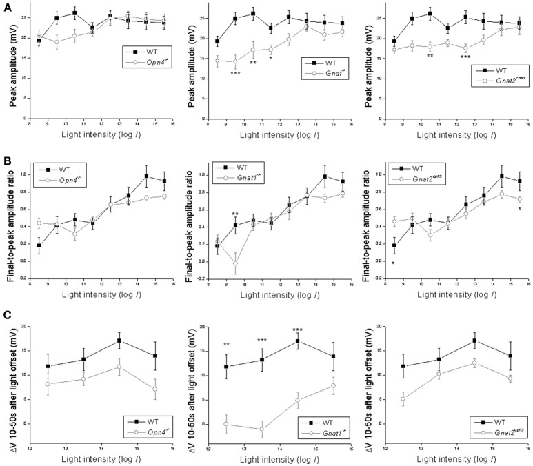
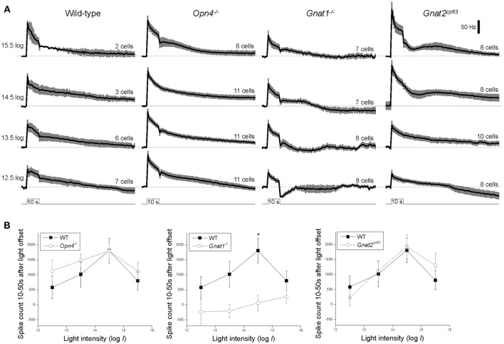
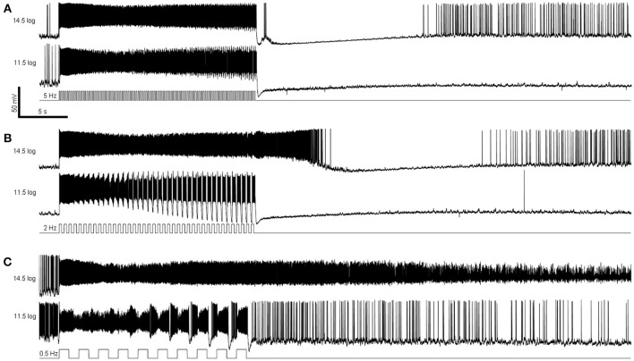
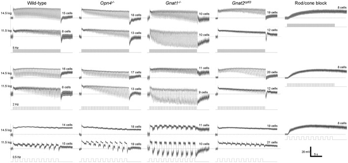
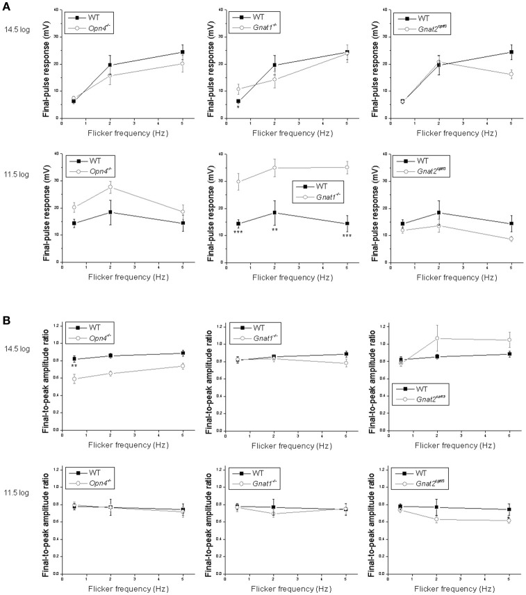
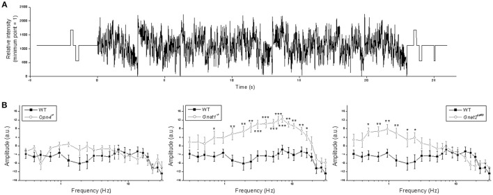
Intrinsically photosensitive retinal ganglion cells (ipRGCs) mediate not only image-forming vision like other ganglion cells, but also non-image-forming physiological responses to light such as pupil constriction and circadian photoentrainment. All ipRGCs respond to light through their endogenous photopigment melanopsin as well as rod/cone-driven synaptic inputs. A major knowledge gap is how melanopsin, rods, and cones differentially drive ipRGC photoresponses and image-forming vision. We whole-cell-recorded from M4-type ipRGCs lacking melanopsin, rod input, or cone input to dissect the roles of each component in ipRGCs' responses to steady and temporally modulated (≥0.3 Hz) lights. We also used a behavioral assay to determine how the elimination of melanopsin, rod, or cone function impacts the optokinetic visual behavior of mice. Results showed that the initial, transient peak in an M4 cell's responses to 10-s light steps arises from rod and cone inputs. Both the sustainability and poststimulus persistence of these light-step responses depend only on rod and/or cone inputs, which is unexpected because these ipRGC photoresponse properties have often been attributed primarily to melanopsin. For temporally varying stimuli, the enhancement of response sustainedness involves melanopsin, whereas stimulus tracking is mediated by rod and cone inputs. Finally, the behavioral assay showed that while all three photoreceptive systems are nearly equally important for contrast sensitivity, only cones and rods contribute to spatial acuity.
内在光敏性视网膜神经节细胞(ipRGCs)不仅像其他神经节细胞一样介导成像视觉,还介导对光的非成像生理反应,如瞳孔收缩和昼夜光同步。所有ipRGCs通过其内源性光色素黑素视蛋白以及视杆/视锥驱动的突触输入对光作出反应。一个主要的知识空白是黑素视蛋白、视杆和视锥如何差异地驱动ipRGC的光反应和成像视觉。我们从缺乏黑素视蛋白、视杆输入或视锥输入的M4型ipRGCs进行全细胞记录,以剖析每个成分在ipRGCs对稳定和时间调制(≥0.3Hz)光反应中的作用。我们还使用行为测定法来确定消除黑素视蛋白、视杆或视锥功能如何影响小鼠的视动视觉行为。结果表明,M4细胞对10秒光刺激的初始瞬态峰值来自视杆和视锥输入。这些光刺激反应的持续性和刺激后持续性仅取决于视杆和/或视锥输入,这是出乎意料的,因为这些ipRGC光反应特性通常主要归因于黑素视蛋白。对于随时间变化的刺激,反应持续性的增强涉及黑素视蛋白,而刺激跟踪由视杆和视锥输入介导。最后,行为测定表明,虽然所有三种光感受系统对对比敏感度几乎同样重要,但只有视锥和视杆对空间敏锐度有贡献。