Chen Jun, Liu Jiapeng, Zeng Jiao, Wu Shan, Ren Jun
Department of Neurology, Affiliated Hospital of Guizhou Medical University, Guiyang, Guizhou Province, China.
Department of Neurology, Lianshui County People's Hospital of Jiangsu Province, Huaian, Jiangsu Province, China.
J Med Ultrasound. 2018 Apr-Jun;26(2):85-89. doi: 10.4103/JMU.JMU_6_17. Epub 2018 May 7.
High-resolution ultrasonography (HRUS) has been used recently to characterize median and ulnar nerves but is seldom used to characterize the lower extremity nerves. The reference standard for normal the lower extremity nerves has not been established. Thus, this study measured the cross-sectional areas (CSAs) of the sciatic nerve of 200 healthy male or female volunteers, aged 18-80 using HRUS. These data provide basic clinical data for the use of high-resolution ultrasound for the future diagnosis, treatment, and prognostic evaluation of peripheral neuropathies.
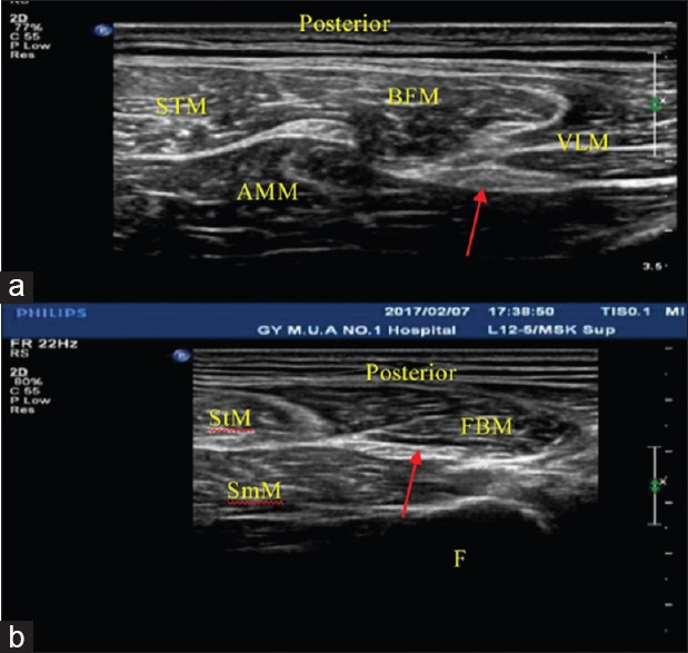
Two hundred healthy volunteers with 400 lower extremities were studied with HRUS. According to their age, the subjects were assigned to young group (18-30 years, = 75), middle group. (31-60 years, = 70), and old group(61-80 year, = 55). Age, sex, height, weight were recorded and CSAs of sciatic nerve were obtained at every predetermined sites.
The mean CSAs of sciatic nerves at GS and MGPF were 0.527 ± 0.028 cm and 0.444 ± 0.026 cm respectively. Pearson's correlation analysis showed that the mean CSAs were correlated with height and weight. There was no difference in mean CSAs among the three groups ( > 0.05). Women had smaller CSAs of the normal Sciatic nerves than men in two measuring sites (GS, MGPF) ( < 0.05).
Peripheral nerve ultrasonography is a reliable and reproducible diagnostic method in the hands of experienced examiners. Normal values for the sciatic nerve nerves are provided by our study. Thus, reference values of Sciatic nerve CSA of the lower extremity can facilitate the analysis of abnormal nerve conditions.
高分辨率超声检查(HRUS)最近已用于描述正中神经和尺神经的特征,但很少用于描述下肢神经的特征。正常下肢神经的参考标准尚未确立。因此,本研究使用HRUS测量了200名年龄在18 - 80岁的健康男性或女性志愿者坐骨神经的横截面积(CSA)。这些数据为未来使用高分辨率超声诊断、治疗和评估周围神经病变提供了基础临床数据。
对200名有400条下肢的健康志愿者进行HRUS检查。根据年龄,将受试者分为青年组(18 - 30岁,n = 75)、中年组(31 - 60岁,n = 70)和老年组(61 - 80岁,n = 55)。记录年龄、性别、身高、体重,并在每个预定部位获取坐骨神经的CSA。
坐骨神经在坐骨结节(GS)和股骨大转子平面(MGPF)处的平均CSA分别为0.527±0.028cm²和0.444±0.026cm²。Pearson相关性分析表明,平均CSA与身高和体重相关。三组之间的平均CSA无差异(P>0.05)。在两个测量部位(GS、MGPF),女性正常坐骨神经的CSA小于男性(P<0.05)。
在经验丰富的检查者手中,周围神经超声检查是一种可靠且可重复的诊断方法。我们的研究提供了坐骨神经的正常值。因此,下肢坐骨神经CSA的参考值有助于分析异常神经情况。