Stenkrona Per, Matheson Granville J, Cervenka Simon, Sigray Pontus Plavén, Halldin Christer, Farde Lars
Department of Clinical Neuroscience, Centre for Psychiatry Research, Karolinska University Hospital, Karolinska Institutet, R5:02, S-171 76, Stockholm, Sweden.
Stockholm County Council, Stockholm, Sweden.
EJNMMI Res. 2018 Aug 2;8(1):74. doi: 10.1186/s13550-018-0416-2.
The D-dopamine receptor radioligand [C]SCH23390 has been frequently used in PET studies. In drug-naïve patients with schizophrenia, the findings have been inconsistent, with decreases, increases, and no change in the frontal cortex D-dopamine receptors. While these discrepancies are likely primarily due to a lack of statistical power in these studies, we speculated that an additional explanation may be the differences due to methods of image analysis between studies, affecting reliability as well as bias between groups.
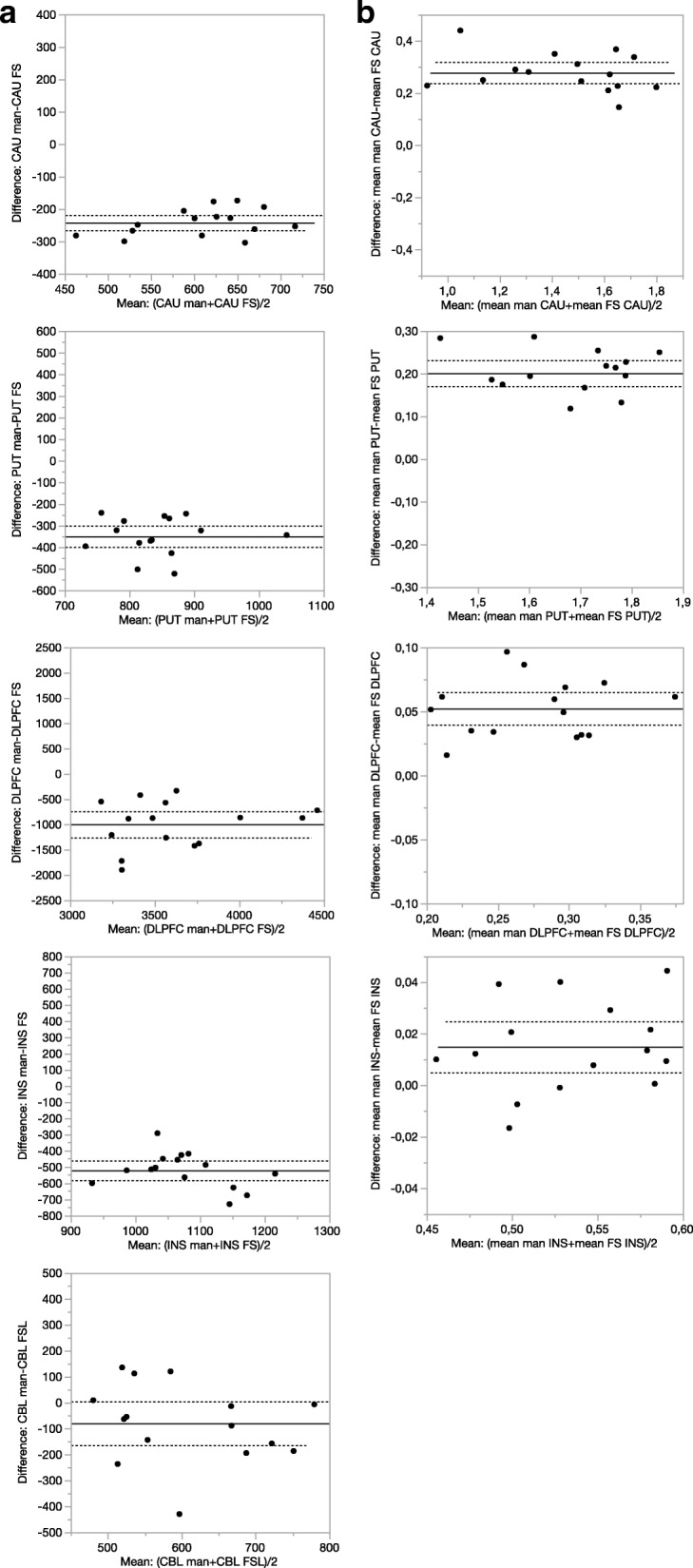
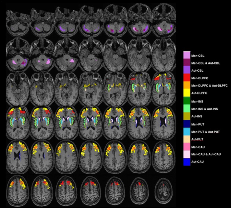
Fifteen healthy subjects underwent two PET measurements with [C]SCH23390 on the same day. The binding potential (BP) was compared using a 95% confidence interval following manual and automated delineation of a region of interest (ROI) as well as with and without frame-by-frame realignment.
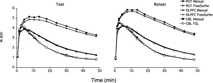
Automated target region delineation produced lower BP values, while automated delineation of the reference region yielded higher BP values. However, no significant differences were observed for repeatability using automated and manual delineation methods. Frame-by-frame realignment generated higher BP values and improved repeatability.
The results suggest that the choice of ROI delineation method is not an important factor for reliability, whereas the improved results following movement correction confirm its importance in PET image analysis. Realignment is therefore especially important for measurements in patient populations such as schizophrenia or Parkinson's disease, where motion artifacts may be more prevalent.
D - 多巴胺受体放射性配体[C]SCH23390已在正电子发射断层扫描(PET)研究中频繁使用。在未服用过药物的精神分裂症患者中,研究结果并不一致,额叶皮质D - 多巴胺受体出现了减少、增加以及无变化等情况。虽然这些差异可能主要是由于这些研究缺乏统计效力,但我们推测另一种解释可能是研究之间图像分析方法的差异,这会影响可靠性以及组间偏差。
15名健康受试者在同一天接受了两次使用[C]SCH23390的PET测量。在手动和自动勾勒感兴趣区域(ROI)以及有无逐帧重新对齐的情况下,使用95%置信区间比较结合潜力(BP)。
自动勾勒目标区域产生较低的BP值,而自动勾勒参考区域产生较高的BP值。然而,使用自动和手动勾勒方法在重复性方面未观察到显著差异。逐帧重新对齐产生较高的BP值并提高了重复性。
结果表明,ROI勾勒方法的选择并非影响可靠性的重要因素,而运动校正后结果的改善证实了其在PET图像分析中的重要性。因此,重新对齐对于精神分裂症或帕金森病等患者群体的测量尤为重要,因为在这些群体中运动伪影可能更为普遍。