Cardoso Miguel, Dos Anjos Pires Maria, Correlo Vitor, Reis Rui, Paulo Manuel, Viegas Carlos
University of Trás-os-Montes e Alto Douro, School of Agrarian and Veterinary Sciences, Department of Veterinary Sciences, Quinta de Prados, Vila Real, Portugal.
Health Sciences Institute of Universidade Católica Portuguesa, Department of Endodontics; Estrada da Circunvalação, Viseu, Portugal.
Iran Endod J. 2018 Summer;13(3):323-330. doi: 10.22037/iej.v13i3.19890.
Biodentine has been scarcely studied as a furcation perforation (FP) repair material, mostly by methodologies. This animal study aimed to compare the histological responses, radiographic, and micro-computed tomographic (micro-CT) outcomes after FP repair with Biodentine or ProRoot MTA (MTA) in dogs' teeth.
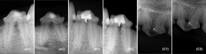
Fifty teeth from five dogs were divided into 4 groups: MTA (=20, FP repaired with ProRoot MTA), BDT (=20, FP repaired with Biodentine), PC (=5, positive control, FP without repair) and NC (=5, negative control, without perforation). The animals were euthanized after 4 months. Histological assessment included inflammatory cell infiltration, hard tissue resorption, hard tissue repair, and cement repair in the furcation area. Immediate postoperative and 4 months follow-up radiographs were compared for radiolucency in the furcation region. The volume of extruded material was quantified using micro-CT images.
The tested materials showed equivalent radiographic response, together with similar hard tissue resorption and repair but, BDT group showed significantly less inflammation, lower volume of extruded material and higher cement repair than MTA group.
The outcomes of this study, taken together with other favorable results in literature, are highly suggestive that Biodentine is a promising biomaterial to be used for FP repair.
作为一种根分叉穿孔(FP)修复材料,生物陶瓷几乎没有被研究过,主要是通过方法学进行研究。本动物研究旨在比较在犬牙中使用生物陶瓷或ProRoot MTA(MTA)修复FP后的组织学反应、影像学和微计算机断层扫描(micro-CT)结果。
将五只犬的50颗牙齿分为4组:MTA组(=20颗,用ProRoot MTA修复FP)、BDT组(=20颗,用生物陶瓷修复FP)、PC组(=5颗,阳性对照,未修复FP)和NC组(=5颗,阴性对照,无穿孔)。4个月后对动物实施安乐死。组织学评估包括根分叉区域的炎性细胞浸润、硬组织吸收、硬组织修复和牙骨质修复。比较术后即刻和4个月随访时根分叉区域的X线透射影像。使用micro-CT图像对挤出材料的体积进行定量分析。
受试材料显示出相同的影像学反应,硬组织吸收和修复情况相似,但BDT组的炎症明显少于MTA组,挤出材料的体积更小,牙骨质修复更高。
本研究结果与文献中的其他有利结果一起,强烈表明生物陶瓷是一种有前途的用于FP修复的生物材料。