Sassi Francesca, Buondonno Ilaria, Luppi Chiara, Spertino Elena, Stratta Emanuela, Di Stefano Marco, Ravazzoli Marco, Isaia Gianluca, Trento Marina, Passera Pietro, Porta Massimo, Isaia Giovanni Carlo, D'Amelio Patrizia
Department of Medical Science, Gerontology and Bone Metabolic Diseases, University of Torino, Corso Bramante 88/90, 10126, Torino, Italy.
Geriatric Division, University of Turin, San Luigi Gonzaga Hospital, Orbassano, Turin, Italy.
BMC Endocr Disord. 2018 Aug 8;18(1):55. doi: 10.1186/s12902-018-0283-x.
Here we study the effect of type 2 diabetes (T2DM) on bone cell precursors, turnover and cytokines involved in the control of bone cell formation and activity.
We enrolled in the study 21 T2DM women and 21 non diabetic controls matched for age and body mass index (BMI). In each subject we measured bone cell precursors, Receptor Activator of Nuclear Factor κB (RANKL), Osteoprotegerin (OPG), Sclerostin (SCL) and Dickoppf-1 (DKK-1) as cytokines involved in the control of osteoblast and osteoclast formation and activity, bone density (BMD) and quality trough trabecular bone score (TBS) and bone turnover. T2DM patients and controls were compared for the analyzed variables by one way ANOVA for Gaussian ones and by Mann-Whitney or Kruskal-Wallis test for non-Gaussian variables.
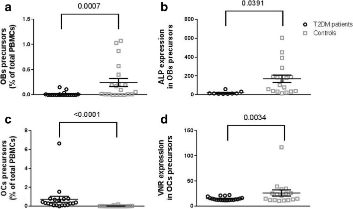
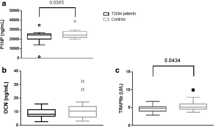
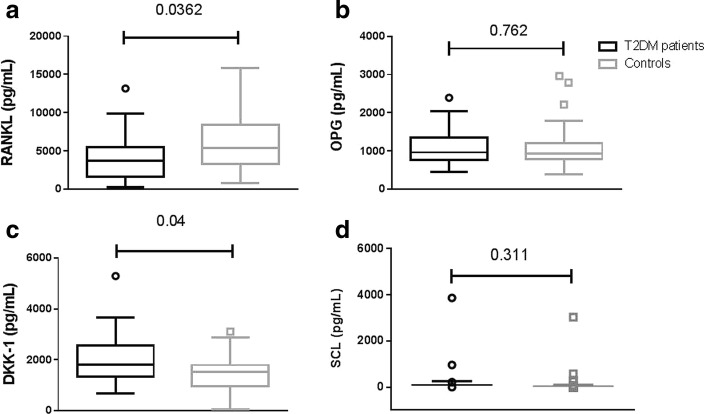
RANKL was decreased and DKK-1 increased in T2DM. Accordingly, patients with T2DM have lower bone turnover compared to controls. BMD and TBS were not significantly different from healthy controls. Bone precursor cells were more immature in T2DM. However the number of osteoclast precursors was increased and that of osteoblasts decreased.
Patients with T2DM have more immature bone cells precursors, with increased number of osteoclasts and decreased osteoblasts, confirming low bone turnover and reduced cytokines such as RANKL and DKK-1. BMD and TBS are not significantly altered in T2DM although, in contrast with other studies, this may be due to the match of patients and controls for BMI rather than age.
在此我们研究2型糖尿病(T2DM)对骨细胞前体、骨转换以及参与骨细胞形成和活性控制的细胞因子的影响。
我们招募了21名T2DM女性患者和21名年龄及体重指数(BMI)匹配的非糖尿病对照者。在每个受试者中,我们测量了骨细胞前体、核因子κB受体激活剂(RANKL)、骨保护素(OPG)、硬化蛋白(SCL)和Dickkopf-1(DKK-1),这些细胞因子参与成骨细胞和破骨细胞的形成及活性控制、骨密度(BMD)以及通过骨小梁骨评分(TBS)和骨转换来评估的骨质量。通过单因素方差分析比较T2DM患者和对照者的高斯分布变量,对于非高斯分布变量则采用Mann-Whitney或Kruskal-Wallis检验来分析变量。
T2DM患者的RANKL降低而DKK-1升高。相应地,与对照组相比,T2DM患者的骨转换较低。BMD和TBS与健康对照组无显著差异。T2DM患者的骨前体细胞更不成熟。然而,破骨细胞前体数量增加而成骨细胞数量减少。
T2DM患者的骨细胞前体更不成熟,破骨细胞数量增加而成骨细胞数量减少,证实骨转换低且RANKL和DKK-1等细胞因子减少。尽管与其他研究不同,T2DM患者的BMD和TBS未显著改变,这可能是由于患者和对照者的BMI匹配而非年龄匹配所致。