Department of Orthodontics and Dentofacial Orthopaedics, Faculty of Dental Medicine, Damascus University, AlMazzah Street, Damascus, Syria.
Prog Orthod. 2018 Aug 13;19(1):29. doi: 10.1186/s40510-018-0232-2.
To investigate the relationship between the morphological maturation stages of the midpalatal suture and its bone densities.
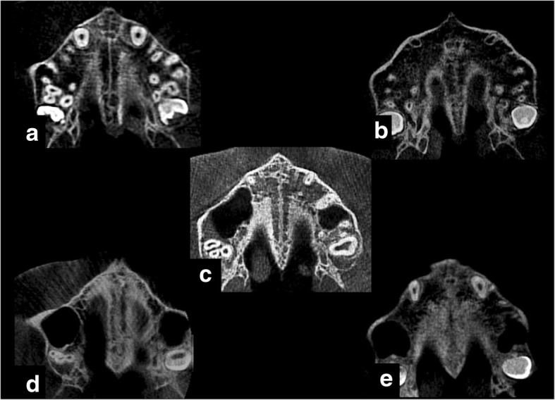
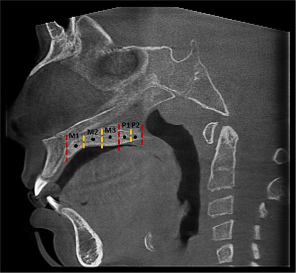
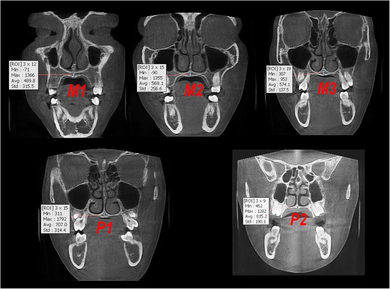
The sample consisted of 91 subjects aged 8-18 years who underwent cone beam computed tomography. All images were examined to classify morphological maturation of the midpalatal suture to five groups according to Angelieri et al. Bone density of the midpalatal suture was measured at the maxillary and palatal regions. Kruskal-Wallis and Mann-Whitney U tests were used to analyze the difference between groups.
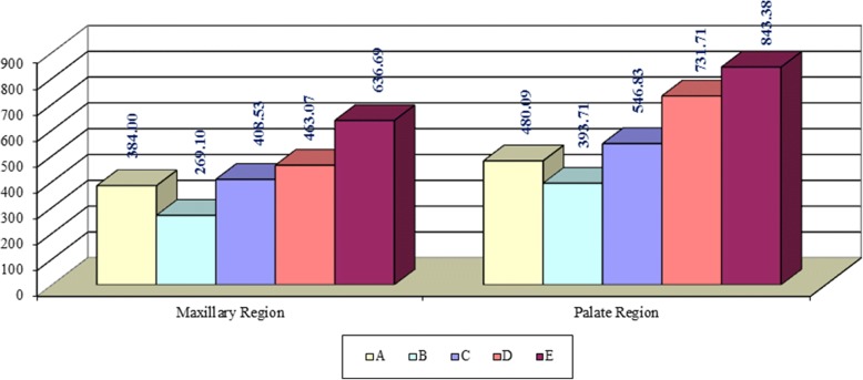
Bone density of the midpalatal suture was significantly higher in the palatal region in E stage and in the maxillary region in D and E stages.
It is concluded that the change in bone density of the midpalatal suture between the morphological maturation stages supports their reliability in clinical application.
研究中隔骨缝形态成熟阶段与其骨密度的关系。
该样本包括 91 名 8-18 岁的受试者,他们接受了锥形束计算机断层扫描。根据 Angelieri 等人的标准,所有图像均进行了检查,以将中隔骨缝的形态成熟分为 5 组。在腭部和上颌区测量中隔骨缝的骨密度。使用 Kruskal-Wallis 和 Mann-Whitney U 检验分析组间差异。
E 期时中隔骨缝的腭部骨密度显著较高,D 期和 E 期时上颌骨骨密度显著较高。
可以得出结论,中隔骨缝形态成熟阶段之间骨密度的变化支持其在临床应用中的可靠性。