Leibniz Research Centre for Working Environment and Human Factors at the Technical University Dortmund, Dortmund, Germany.
Department of Forensic Medicine and Toxicology, Faculty of Veterinary Medicine, South Valley University, Qena, Egypt.
Hepatology. 2019 Feb;69(2):666-683. doi: 10.1002/hep.30213. Epub 2018 Nov 19.
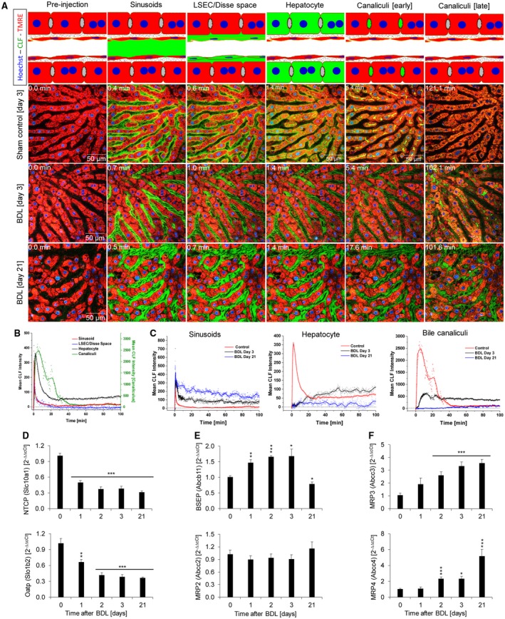
Bile duct ligation (BDL) is an experimental procedure that mimics obstructive cholestatic disease. One of the early consequences of BDL in rodents is the appearance of so-called bile infarcts that correspond to Charcot-Gombault necrosis in human cholestasis. The mechanisms causing bile infarcts and their pathophysiological relevance are unclear. Therefore, intravital two photon-based imaging of BDL mice was performed with fluorescent bile salts (BS) and non-BS organic anion analogues. Key findings were followed up by matrix-assisted laser desorption ionization imaging, clinical chemistry, immunostaining, and gene expression analyses. In the acute phase, 1-3 days after BDL, BS concentrations in bile increased and single-cell bile microinfarcts occurred in dispersed hepatocytes throughout the liver caused by the rupture of the apical hepatocyte membrane. This rupture occurred after loss of mitochondrial membrane potential, followed by entry of bile, cell death, and a "domino effect" of further death events of neighboring hepatocytes. Bile infarcts provided a trans-epithelial shunt between bile canaliculi and sinusoids by which bile constituents leaked into blood. In the chronic phase, ≥21 days after BDL, uptake of BS tracers at the sinusoidal hepatocyte membrane was reduced. This contributes to elevated concentrations of BS in blood and decreased concentrations in the biliary tract. Conclusion: Bile microinfarcts occur in the acute phase after BDL in a limited number of dispersed hepatocytes followed by larger infarcts involving neighboring hepatocytes, and they allow leakage of bile from the BS-overloaded biliary tract into blood, thereby protecting the liver from BS toxicity; in the chronic phase after BDL, reduced sinusoidal BS uptake is a dominant protective factor, and the kidney contributes to the elimination of BS until cholemic nephropathy sets in.
胆管结扎(BDL)是一种模拟阻塞性胆汁淤积疾病的实验程序。BDL 后啮齿动物的早期后果之一是出现所谓的胆汁梗死,这与人类胆汁淤积中的 Charcot-Gombault 坏死相对应。导致胆汁梗死的机制及其病理生理相关性尚不清楚。因此,对 BDL 小鼠进行了基于双光子的活体成像,使用荧光胆汁盐(BS)和非 BS 有机阴离子类似物。主要发现通过基质辅助激光解吸电离成像、临床化学、免疫染色和基因表达分析进行了跟进。在急性阶段,BDL 后 1-3 天,胆汁中的 BS 浓度增加,并且由于顶泌膜破裂,整个肝脏中分散的肝细胞中发生单细胞胆汁微梗死。这种破裂发生在线粒体膜电位丧失之后,随后是胆汁进入、细胞死亡以及相邻肝细胞进一步死亡事件的“多米诺骨牌效应”。胆汁梗死通过上皮细胞间短路为胆汁成分从胆管漏入血液提供了途径。在慢性阶段,BDL 后≥21 天,窦状肝细胞膜上的 BS 示踪剂摄取减少。这导致 BS 在血液中的浓度升高和在胆道中的浓度降低。结论:BDL 后急性阶段,少数分散的肝细胞中发生胆汁微梗死,随后较大的梗死涉及相邻的肝细胞,并且允许 BS 过载的胆道中的胆汁从漏入血液中,从而保护肝脏免受 BS 毒性的侵害;BDL 后慢性阶段,减少的窦状 BS 摄取是一个主要的保护因素,肾脏有助于 BS 的清除,直到出现胆汁性肾病。