• 文献检索
  • 文档翻译
  • 深度研究
  • 学术资讯
  • Suppr Zotero 插件Zotero 插件
  • 邀请有礼
  • 套餐&价格
  • 历史记录
应用&插件
Suppr Zotero 插件Zotero 插件浏览器插件Mac 客户端Windows 客户端微信小程序
定价
高级版会员购买积分包购买API积分包
服务
文献检索文档翻译深度研究API 文档MCP 服务
关于我们
关于 Suppr公司介绍联系我们用户协议隐私条款
关注我们

Suppr 超能文献

核心技术专利:CN118964589B侵权必究
粤ICP备2023148730 号-1Suppr @ 2026

文献检索

告别复杂PubMed语法,用中文像聊天一样搜索,搜遍4000万医学文献。AI智能推荐,让科研检索更轻松。

立即免费搜索

文件翻译

保留排版,准确专业,支持PDF/Word/PPT等文件格式,支持 12+语言互译。

免费翻译文档

深度研究

AI帮你快速写综述,25分钟生成高质量综述,智能提取关键信息,辅助科研写作。

立即免费体验

伴有瘤内出血并表现为急性痉挛性四肢瘫的颈段C2至C4神经鞘瘤:一例罕见病例报告

Cervical C2 to C4 schwannoma with intratumoral hemorrhage presenting as acute spastic quadriparesis: A rare case report.

作者信息

Gandhoke C S, Syal S K, Singh D, Batra V, Nallacheruvu Y

机构信息

Department of Neurosurgery, Maulana Azad Medical College, Lok Nayak Jai Prakash Narayan Hospital, Guru Nanak Eye Centre and G. B. Pant Institute of Postgraduate Medical Education and Research (G.I.P.M.E.R.), New Delhi, India.

Department of Paediatrics, Sri Guru Ram Das Institute of Medical Sciences and Research, Amritsar, Punjab, India.

出版信息

Surg Neurol Int. 2018 Jul 24;9:142. doi: 10.4103/sni.sni_171_18. eCollection 2018.

DOI:10.4103/sni.sni_171_18
PMID:30105136
原文链接:https://pmc.ncbi.nlm.nih.gov/articles/PMC6069368/
Abstract

BACKGROUND

Spinal schwannomas are slow growing, benign nerve sheath tumors. These may be asymptomatic or may present as backache with radicular pain, slowly progressive neurological deficits, but rarely with acute spastic quadriparesis attributed to intratumoral hemorrhage.

CASE DESCRIPTION

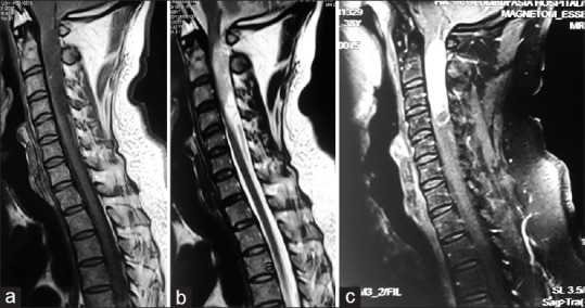
A 38-year-old male presented with the chief complaint of neck pain radiating to the left upper extremity for the last 8 months. On admission, he exhibited diffuse hyper-reflexia but had no motor or sensory deficit. Magnetic resonance imaging showed a solid-cystic intradural extramedullary (IDEM) C2 to C4 mass severely compressing the spinal cord. The same day the patient acutely developed a spastic quadriparesis. Immediately, a partial C2, C3, and C4 laminectomy was performed for tumor excision; within 5 postoperative days, he fully regained neurological function. The final histopathology was consistent with a "schwannoma showing areas of congestion and hemorrhage."

CONCLUSION

Spinal schwannomas rarely present with intratumoral hemorrhage and acute spastic quadriparesis. Immediate operative decompression may lead to excellent postoperative neurological recovery.

摘要

背景

脊髓神经鞘瘤是生长缓慢的良性神经鞘瘤。这些肿瘤可能无症状,或表现为伴有神经根性疼痛的背痛、缓慢进展的神经功能缺损,但很少因肿瘤内出血导致急性痉挛性四肢瘫。

病例描述

一名38岁男性,主要诉求为过去8个月颈部疼痛并向左上肢放射。入院时,他表现为弥漫性反射亢进,但无运动或感觉障碍。磁共振成像显示C2至C4水平有一个实性囊性硬膜内髓外(IDEM)肿块,严重压迫脊髓。同一天,患者急性出现痉挛性四肢瘫。随即进行了C2、C3和C4部分椎板切除术以切除肿瘤;术后5天内,他完全恢复了神经功能。最终组织病理学结果与“显示充血和出血区域的神经鞘瘤”一致。

结论

脊髓神经鞘瘤很少出现肿瘤内出血和急性痉挛性四肢瘫。立即进行手术减压可能导致术后神经功能极佳的恢复情况。

https://cdn.ncbi.nlm.nih.gov/pmc/blobs/240c/6069368/fbd04deaa902/SNI-9-142-g003.jpg
https://cdn.ncbi.nlm.nih.gov/pmc/blobs/240c/6069368/c1f4e10b7469/SNI-9-142-g001.jpg
https://cdn.ncbi.nlm.nih.gov/pmc/blobs/240c/6069368/1a33b074ab3f/SNI-9-142-g002.jpg
https://cdn.ncbi.nlm.nih.gov/pmc/blobs/240c/6069368/fbd04deaa902/SNI-9-142-g003.jpg
https://cdn.ncbi.nlm.nih.gov/pmc/blobs/240c/6069368/c1f4e10b7469/SNI-9-142-g001.jpg
https://cdn.ncbi.nlm.nih.gov/pmc/blobs/240c/6069368/1a33b074ab3f/SNI-9-142-g002.jpg
https://cdn.ncbi.nlm.nih.gov/pmc/blobs/240c/6069368/fbd04deaa902/SNI-9-142-g003.jpg

相似文献

1
Cervical C2 to C4 schwannoma with intratumoral hemorrhage presenting as acute spastic quadriparesis: A rare case report.伴有瘤内出血并表现为急性痉挛性四肢瘫的颈段C2至C4神经鞘瘤:一例罕见病例报告
Surg Neurol Int. 2018 Jul 24;9:142. doi: 10.4103/sni.sni_171_18. eCollection 2018.
2
Intratumoral Hemorrhage of the Cervical Spinal Schwannoma Presenting: Acute Quadriparesis.颈段脊髓神经鞘瘤瘤内出血表现为急性四肢瘫。
Brain Tumor Res Treat. 2019 Oct;7(2):160-163. doi: 10.14791/btrt.2019.7.e34.
3
Acute hemorrhage within intradural extramedullary schwannoma in cervical spine presenting with quadriparesis.颈椎硬膜内髓外神经鞘瘤内急性出血伴四肢瘫。
J Craniovertebr Junction Spine. 2015 Apr-Jun;6(2):83-5. doi: 10.4103/0974-8237.156069.
4
Totally cystic intradural schwannoma in thoracic region.胸段完全囊性硬膜内神经鞘瘤
Asian J Neurosurg. 2017 Jan-Mar;12(1):131-133. doi: 10.4103/1793-5482.145570.
5
Cervical schwannoma with acute worsening and intratumoral hemorrhage.伴有急性病情恶化及瘤内出血的颈椎神经鞘瘤
Surg Neurol Int. 2021 Aug 16;12:409. doi: 10.25259/SNI_623_2021. eCollection 2021.
6
Acute paraplegia due to thoracolumbar schwannoma following trauma: A case report and literature review.创伤后胸段及腰段神经鞘瘤所致急性截瘫:一例报告并文献复习
Mol Clin Oncol. 2021 Oct;15(4):204. doi: 10.3892/mco.2021.2366. Epub 2021 Aug 8.
7
Solitary osteochondroma of the cervical spine presenting with quadriparesis and hand contracture.颈椎孤立性骨软骨瘤伴四肢瘫和手部挛缩。
Surg Neurol Int. 2020 Mar 21;11:51. doi: 10.25259/SNI_3_2020. eCollection 2020.
8
Intramedullary and intratumoral hemorrhage in spinal hemangioblastoma: Case report and review of literature.脊髓血管母细胞瘤的髓内及瘤内出血:病例报告及文献复习
Surg Neurol Int. 2018 Dec 4;9:250. doi: 10.4103/sni.sni_344_18. eCollection 2018.
9
Giant Invasive Intradural Extramedullary Lumbar Schwannoma: A Case Report and Literature Review.巨大侵袭性硬脊膜内髓外腰椎神经鞘瘤:病例报告及文献综述
Cureus. 2023 Jun 20;15(6):e40708. doi: 10.7759/cureus.40708. eCollection 2023 Jun.
10
Successful excision of intradural extramedullary plexiform T1-T4 schwannoma.成功切除硬脊膜内髓外T1 - T4节段的丛状神经鞘瘤。
Surg Neurol Int. 2024 Sep 6;15:321. doi: 10.25259/SNI_617_2024. eCollection 2024.

引用本文的文献

1
Spinal schwannoma presenting with intraspinal hematoma: a case report and review of the literature.脊髓神经鞘瘤伴脊髓内血肿:一例报告并文献复习
J Med Case Rep. 2024 Dec 21;18(1):633. doi: 10.1186/s13256-024-04968-4.
2
Intradural cystic schwannomas of the spine: A case-based systematic review of an unusual tumor.脊柱硬膜内囊性神经鞘瘤:基于病例的罕见肿瘤系统综述
Brain Spine. 2024 May 28;4:102843. doi: 10.1016/j.bas.2024.102843. eCollection 2024.
3
Acute Neurological Aggravation Caused by Intratumoral Hemorrhage of a Cervical Dumbbell Schwannoma: Report of a Rare Case and Literature Review.

本文引用的文献

1
Giant cystic intradural schwannoma in the lumbosacral region: a case report.腰骶部巨大囊性硬膜内神经鞘瘤:病例报告
J Orthop Surg (Hong Kong). 2008 Apr;16(1):102-6. doi: 10.1177/230949900801600124.
2
Cystic lumbar nerve sheath tumours: MRI features in five patients.囊性腰神经鞘瘤:5例患者的MRI特征
Australas Radiol. 2001 May;45(2):123-7. doi: 10.1046/j.1440-1673.2001.00891.x.
3
MRI of intramedullary spinal schwannomas: case report and review of the literature.脊髓髓内神经鞘瘤的磁共振成像:病例报告及文献复习
颈哑铃型神经鞘瘤瘤内出血导致的急性神经功能恶化:1例罕见病例报告及文献复习
Cureus. 2023 Feb 6;15(2):e34682. doi: 10.7759/cureus.34682. eCollection 2023 Feb.
4
Clinical Effect of Laminectomy with Lateral Mass Screw Fixation in Treating Cervical Schwannoma: A Retrospective Study.经侧块螺钉固定椎板切除术治疗颈椎神经鞘瘤的临床效果:一项回顾性研究。
Biomed Res Int. 2022 Apr 29;2022:8512374. doi: 10.1155/2022/8512374. eCollection 2022.
5
Hemorrhagic Schwannoma of the Cauda Equina: Case Report and Review of the Literature.马尾神经出血性神经鞘瘤:病例报告及文献复习
NMC Case Rep J. 2021 Jul 9;8(1):377-385. doi: 10.2176/nmccrj.cr.2020-0186. eCollection 2021.
6
Cervical schwannoma with acute worsening and intratumoral hemorrhage.伴有急性病情恶化及瘤内出血的颈椎神经鞘瘤
Surg Neurol Int. 2021 Aug 16;12:409. doi: 10.25259/SNI_623_2021. eCollection 2021.
Neuroradiology. 2000 Apr;42(4):275-9. doi: 10.1007/s002340050885.
4
Spinal subarachnoid hemorrhage attributable to schwannoma of the cauda equina.
Surg Neurol. 1999 Apr;51(4):373-5. doi: 10.1016/s0090-3019(98)00046-9.
5
Spinal subarachnoid haemorrhage from an "ancient" schwannoma of the cervical spine.
Br J Neurosurg. 1993;7(5):557-9. doi: 10.3109/02688699308995079.
6
Subarachnoid hemorrhage from malignant schwannoma of cauda equina.
Tex Med. 1973 Oct;69(10):101-4.
7
Subarachnoid hemorrhage due to a spinal neurinoma.
Surg Neurol. 1985 Aug;24(2):187-90. doi: 10.1016/0090-3019(85)90184-3.
8
MR imaging of spinal neurinomas with pathological correlation.
J Comput Assist Tomogr. 1990 Mar-Apr;14(2):250-4. doi: 10.1097/00004728-199003000-00017.
9
Intradural schwannomas of the spine: MR findings with emphasis on contrast-enhancement characteristics.脊柱硬膜内神经鞘瘤:磁共振成像表现,重点关注增强特征
AJR Am J Roentgenol. 1992 Jun;158(6):1347-50. doi: 10.2214/ajr.158.6.1590138.
10
Subarachnoid hemorrhage and papilledema due to a cervical neurilemmoma. Case report.
J Neurosurg. 1978 Jun;48(6):1015-8. doi: 10.3171/jns.1978.48.6.1015.