Department of Geriatric Cardiovascular Medicine, Sichuan Academy of Medical Sciences and Sichuan Provincial People's Hospital, Chengdu, Sichuan 610072, P.R. China.
Department of Cardiovascular Medicine, The Affiliated Hospital of Southwest Medical University, Luzhou, Sichuan 646000, P.R. China.
Mol Med Rep. 2018 Oct;18(4):3641-3648. doi: 10.3892/mmr.2018.9354. Epub 2018 Aug 3.
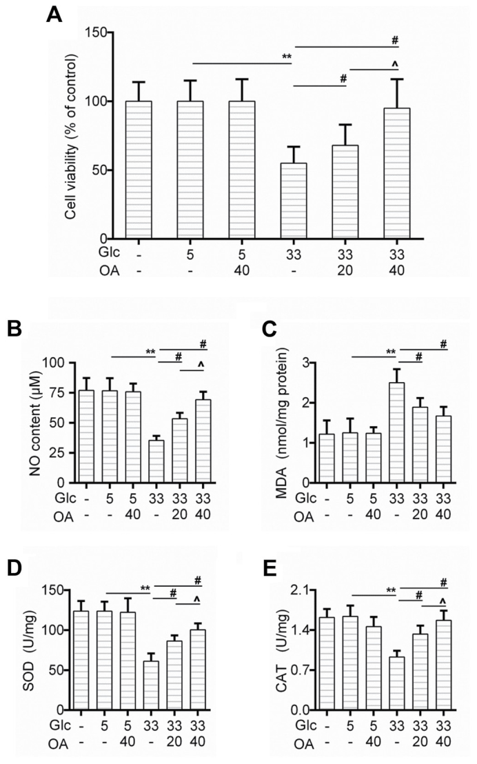
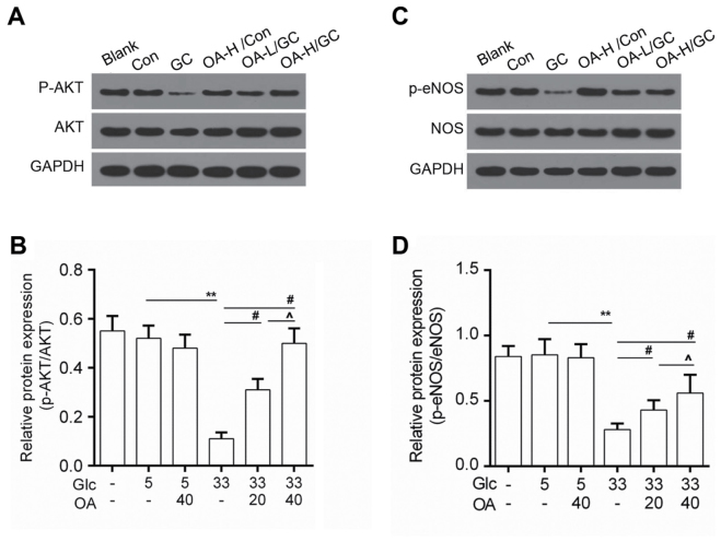
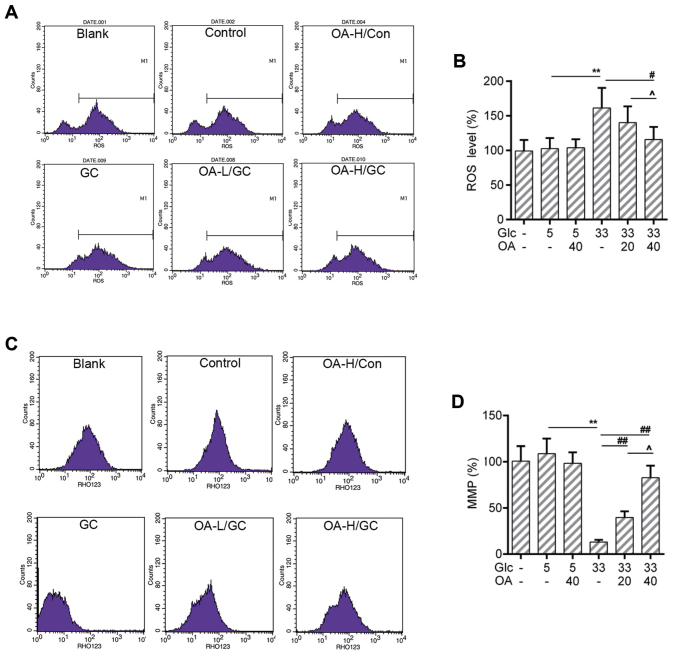
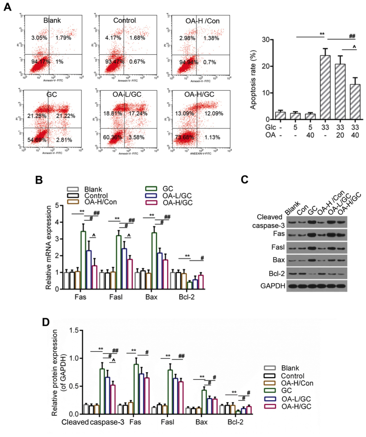
Oxidative injury of vascular endothelial cells in the initial event of atherosclerosis (AS) in diabetes was assessed in the present study. The antioxidant effect of oleanolic acid (OA) has attracted much attention. In the present study the potential effects of OA on human umbilical vein endothelial cells (HUVECs) were investigated. Cell viability was examined using the CCK‑8 assay. The activity of oxidative stress parameters was determined using commercial kits. Flow cytometry analysis was performed to detect the level of reactive oxygen species (ROS), mitochondrial membrane potential (MMP) and cell apoptosis. The expression levels of target genes and proteins were examined by reverse transcription‑quantitative polymerase chain reaction (RT‑qPCR) and western blot analysis. It was indicated that cell viability that was suppressed by high glucose was increased by the pretreatment of OA, and nitric oxide (NO) generation, the activities of superoxide dismutase (SOD) and catalase (CAT) were recovered by OA. By contrast, it was observed that OA decreased the MDA content. Notably, the pretreatment of OA alleviated mitochondria damage by reducing the level of ROS and maintaining MMP. In addition, apoptosis that was caused by high glucose was reduced by OA. Pro‑apoptotic genes (caspase‑3, Fas, Fasl) and anti‑apoptotic gene (Bcl‑2) expression levels were decreased and increased in the OA groups, respectively. Furthermore, the activity of AKT/endothelial nitric oxide synthase (eNOS) signaling was elevated by OA. Taken together, it was suggested that OA could protect against oxidative stress‑induced apoptosis of HUVECs, which was associated with AKT/eNOS signaling pathway.
本研究旨在评估糖尿病患者动脉粥样硬化(AS)初始事件中血管内皮细胞的氧化损伤。齐墩果酸(OA)的抗氧化作用引起了广泛关注。本研究旨在探讨 OA 对人脐静脉内皮细胞(HUVEC)的潜在作用。通过 CCK-8 测定法检测细胞活力。使用商业试剂盒测定氧化应激参数的活性。通过流式细胞术分析检测活性氧(ROS)、线粒体膜电位(MMP)和细胞凋亡水平。通过反转录-定量聚合酶链反应(RT-qPCR)和蛋白质印迹分析检测靶基因和蛋白质的表达水平。结果表明,高葡萄糖抑制的细胞活力通过 OA 的预处理而增加,并且 NO 生成、超氧化物歧化酶(SOD)和过氧化氢酶(CAT)的活性被 OA 恢复。相比之下,OA 降低了 MDA 含量。值得注意的是,OA 通过降低 ROS 水平和维持 MMP 减轻了线粒体损伤。此外,OA 减少了由高葡萄糖引起的细胞凋亡。促凋亡基因(caspase-3、Fas、Fasl)和抗凋亡基因(Bcl-2)的表达水平在 OA 组中分别降低和升高。此外,OA 增强了 AKT/内皮型一氧化氮合酶(eNOS)信号通路的活性。综上所述,OA 可防止氧化应激诱导的 HUVEC 凋亡,这与 AKT/eNOS 信号通路有关。