• 文献检索
  • 文档翻译
  • 深度研究
  • 学术资讯
  • Suppr Zotero 插件Zotero 插件
  • 邀请有礼
  • 套餐&价格
  • 历史记录
应用&插件
Suppr Zotero 插件Zotero 插件浏览器插件Mac 客户端Windows 客户端微信小程序
定价
高级版会员购买积分包购买API积分包
服务
文献检索文档翻译深度研究API 文档MCP 服务
关于我们
关于 Suppr公司介绍联系我们用户协议隐私条款
关注我们

Suppr 超能文献

核心技术专利:CN118964589B侵权必究
粤ICP备2023148730 号-1Suppr @ 2026

文献检索

告别复杂PubMed语法,用中文像聊天一样搜索,搜遍4000万医学文献。AI智能推荐,让科研检索更轻松。

立即免费搜索

文件翻译

保留排版,准确专业,支持PDF/Word/PPT等文件格式,支持 12+语言互译。

免费翻译文档

深度研究

AI帮你快速写综述,25分钟生成高质量综述,智能提取关键信息,辅助科研写作。

立即免费体验

FluoEM,用于三维电子显微镜数据中远距离连接组学的轴突虚拟标记。

FluoEM, virtual labeling of axons in three-dimensional electron microscopy data for long-range connectomics.

机构信息

Department of Connectomics, Max Planck Institute for Brain Research, Frankfurt, Germany.

Donders Institute, Faculty of Science, Radboud University, Nijmegen, Netherlands.

出版信息

Elife. 2018 Aug 14;7:e38976. doi: 10.7554/eLife.38976.

DOI:10.7554/eLife.38976
PMID:30106377
原文链接:https://pmc.ncbi.nlm.nih.gov/articles/PMC6158011/
Abstract

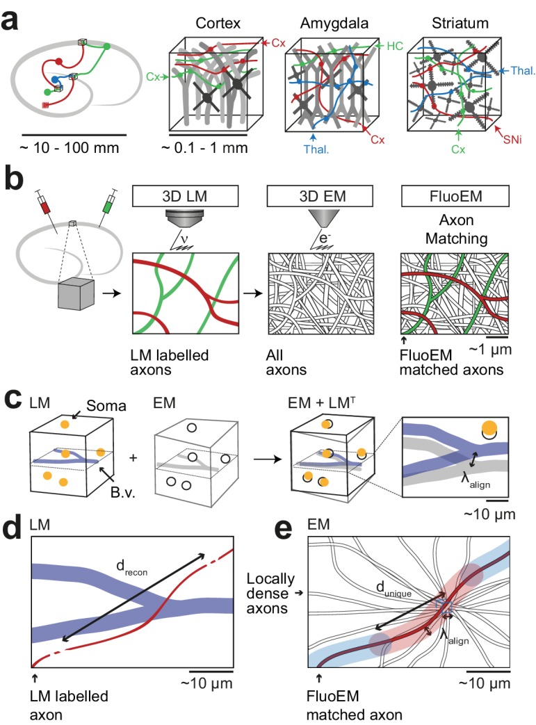
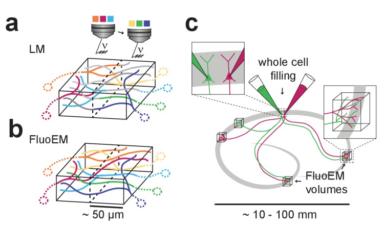
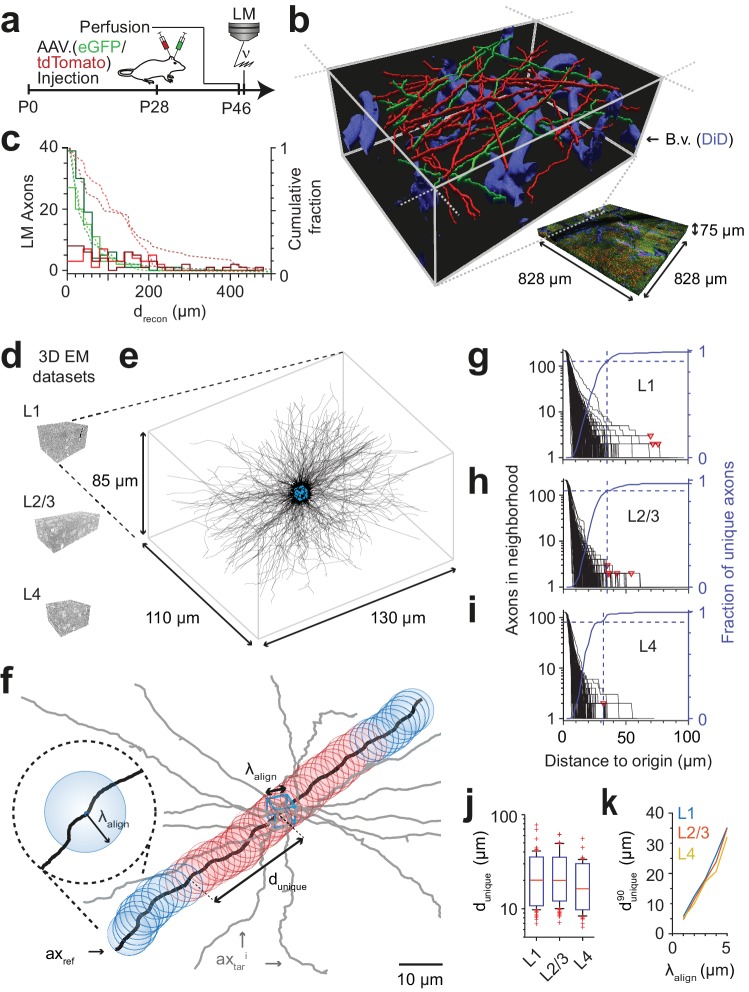
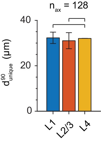
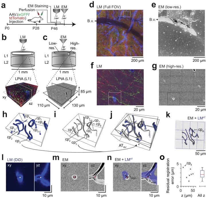
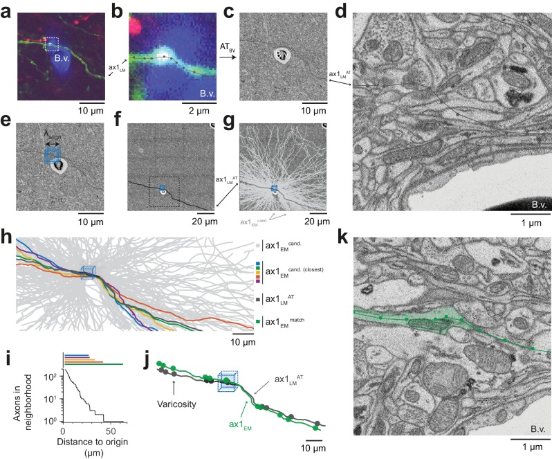
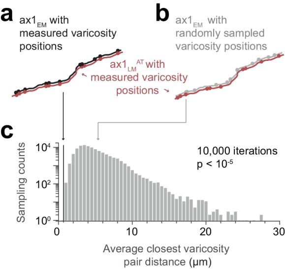
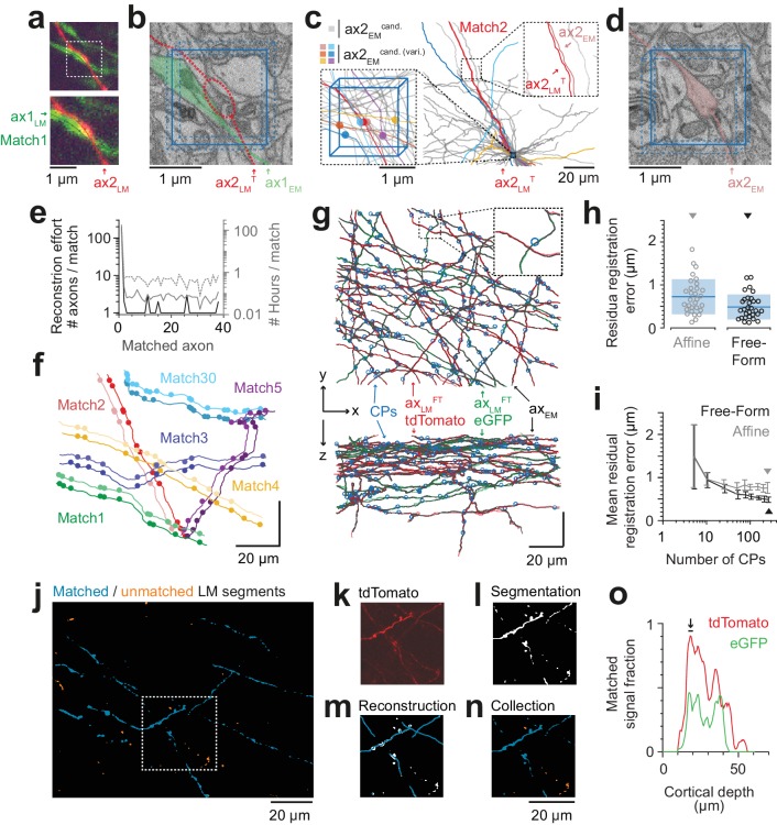
The labeling and identification of long-range axonal inputs from multiple sources within densely reconstructed electron microscopy (EM) datasets from mammalian brains has been notoriously difficult because of the limited color label space of EM. Here, we report FluoEM for the identification of multi-color fluorescently labeled axons in dense EM data without the need for artificial fiducial marks or chemical label conversion. The approach is based on correlated tissue imaging and computational matching of neurite reconstructions, amounting to a virtual color labeling of axons in dense EM circuit data. We show that the identification of fluorescent light- microscopically (LM) imaged axons in 3D EM data from mouse cortex is faithfully possible as soon as the EM dataset is about 40-50 µm in extent, relying on the unique trajectories of axons in dense mammalian neuropil. The method is exemplified for the identification of long-distance axonal input into layer 1 of the mouse cerebral cortex.

摘要

在哺乳动物大脑的密集重建电子显微镜 (EM) 数据集内,对来自多个来源的长程轴突输入进行标记和识别一直是一个难题,因为 EM 的颜色标签空间有限。在这里,我们报告了 FluoEM,用于在不需要人工基准标记或化学标签转换的情况下,识别密集 EM 数据中的多色荧光标记轴突。该方法基于组织的相关成像和神经突重建的计算匹配,相当于对密集 EM 电路数据中的轴突进行虚拟颜色标记。我们表明,只要 EM 数据集的范围约为 40-50 µm,就可以忠实地识别来自小鼠皮层的 3D EM 数据中荧光显微镜 (LM) 成像的轴突,这依赖于密集哺乳动物神经胶质中轴突的独特轨迹。该方法以识别长距离轴突输入到小鼠大脑皮层第 1 层为例进行了说明。

https://cdn.ncbi.nlm.nih.gov/pmc/blobs/58ca/6158011/a4620d07f0c0/elife-38976-fig7.jpg
https://cdn.ncbi.nlm.nih.gov/pmc/blobs/58ca/6158011/c29b34d67b4e/elife-38976-fig1.jpg
https://cdn.ncbi.nlm.nih.gov/pmc/blobs/58ca/6158011/9f3bf3329a4a/elife-38976-fig2.jpg
https://cdn.ncbi.nlm.nih.gov/pmc/blobs/58ca/6158011/3ada6ca94960/elife-38976-fig2-figsupp1.jpg
https://cdn.ncbi.nlm.nih.gov/pmc/blobs/58ca/6158011/4e514c370574/elife-38976-fig3.jpg
https://cdn.ncbi.nlm.nih.gov/pmc/blobs/58ca/6158011/a1f240328c64/elife-38976-fig4.jpg
https://cdn.ncbi.nlm.nih.gov/pmc/blobs/58ca/6158011/c810f0fb3297/elife-38976-fig4-figsupp1.jpg
https://cdn.ncbi.nlm.nih.gov/pmc/blobs/58ca/6158011/679122cfa54c/elife-38976-fig5.jpg
https://cdn.ncbi.nlm.nih.gov/pmc/blobs/58ca/6158011/de7e7fa1606b/elife-38976-fig5-figsupp1.jpg
https://cdn.ncbi.nlm.nih.gov/pmc/blobs/58ca/6158011/8db715eed683/elife-38976-fig6.jpg
https://cdn.ncbi.nlm.nih.gov/pmc/blobs/58ca/6158011/a4620d07f0c0/elife-38976-fig7.jpg

相似文献

1
FluoEM, virtual labeling of axons in three-dimensional electron microscopy data for long-range connectomics.FluoEM,用于三维电子显微镜数据中远距离连接组学的轴突虚拟标记。
Elife. 2018 Aug 14;7:e38976. doi: 10.7554/eLife.38976.
2
Large-volume en-bloc staining for electron microscopy-based connectomics.用于基于电子显微镜的连接组学的大体积整体染色。
Nat Commun. 2015 Aug 3;6:7923. doi: 10.1038/ncomms8923.
3
Dense connectomic reconstruction in layer 4 of the somatosensory cortex.躯体感觉皮层第 4 层的密集连接组构重建。
Science. 2019 Nov 29;366(6469). doi: 10.1126/science.aay3134. Epub 2019 Oct 24.
4
Semiautomated correlative 3D electron microscopy of in vivo-imaged axons and dendrites.活体内成像轴突和树突的半自动关联三维电子显微镜。
Nat Protoc. 2014;9(6):1354-66. doi: 10.1038/nprot.2014.101. Epub 2014 May 15.
5
A protocol for preparing GFP-labeled neurons previously imaged in vivo and in slice preparations for light and electron microscopic analysis.一种用于制备绿色荧光蛋白(GFP)标记神经元的方案,这些神经元先前已在体内和脑片标本中成像,用于光学和电子显微镜分析。
Nat Protoc. 2009;4(8):1145-56. doi: 10.1038/nprot.2009.114. Epub 2009 Jul 16.
6
Morphological Characterization of a Cortico-cortical relay in the cat sensorimotor cortex.猫感觉运动皮层中皮质-皮质中继的形态学特征
Cereb Cortex. 1997 Mar;7(2):100-9. doi: 10.1093/cercor/7.2.100.
7
Correlative in vivo 2-photon imaging and focused ion beam scanning electron microscopy: 3D analysis of neuronal ultrastructure.体内双光子成像与聚焦离子束扫描电子显微镜的相关性:神经元超微结构的三维分析
Methods Cell Biol. 2014;124:339-61. doi: 10.1016/B978-0-12-801075-4.00016-1.
8
Axonal synapse sorting in medial entorhinal cortex.内侧隔核的轴突突触分类。
Nature. 2017 Sep 28;549(7673):469-475. doi: 10.1038/nature24005. Epub 2017 Sep 20.
9
Genetically targeted 3D visualisation of Drosophila neurons under Electron Microscopy and X-Ray Microscopy using miniSOG.利用 miniSOG 在电子显微镜和 X 射线显微镜下对果蝇神经元进行基因靶向 3D 可视化。
Sci Rep. 2016 Dec 13;6:38863. doi: 10.1038/srep38863.
10
Circuitry and role of substance P-immunoreactive neurons in the primate retina.灵长类视网膜中P物质免疫反应性神经元的回路及作用
J Comp Neurol. 1998 Apr 20;393(4):439-56.

引用本文的文献

1
Diazinon residues levels in farm-gate Brassica oleracea var. acephala of Kimira-Oluch smallholder farm improvement project, Kenya.肯尼亚基米拉 - 奥卢奇小农户农场改良项目中,农场门口羽衣甘蓝上的二嗪农残留水平。
PLoS One. 2025 May 28;20(5):e0310586. doi: 10.1371/journal.pone.0310586. eCollection 2025.
2
Cognitive synaptopathy: synaptic and dendritic spine dysfunction in age-related cognitive disorders.认知性突触病变:年龄相关性认知障碍中的突触和树突棘功能障碍
Front Aging Neurosci. 2024 Oct 3;16:1476909. doi: 10.3389/fnagi.2024.1476909. eCollection 2024.
3
Dense 4D nanoscale reconstruction of living brain tissue.

本文引用的文献

1
High-precision automated reconstruction of neurons with flood-filling networks.基于填充网络的高精度自动化神经元重建。
Nat Methods. 2018 Aug;15(8):605-610. doi: 10.1038/s41592-018-0049-4. Epub 2018 Jul 16.
2
High-quality ultrastructural preservation using cryofixation for 3D electron microscopy of genetically labeled tissues.使用 cryofixation 进行高质量的超微结构保存,用于基因标记组织的 3D 电子显微镜观察。
Elife. 2018 May 11;7:e35524. doi: 10.7554/eLife.35524.
3
Computer assisted detection of axonal bouton structural plasticity in time-lapse images.
活脑组织的密集 4D 纳米尺度重建。
Nat Methods. 2023 Aug;20(8):1256-1265. doi: 10.1038/s41592-023-01936-6. Epub 2023 Jul 10.
4
Reconstructing neural circuits using multiresolution correlated light and electron microscopy.使用多分辨率相关光和电子显微镜重建神经回路。
Front Neural Circuits. 2022 Oct 21;16:753496. doi: 10.3389/fncir.2022.753496. eCollection 2022.
5
A new form of axonal pathology in a spinal model of neuromyelitis optica.视神经脊髓炎模型中新形式的轴突病理学。
Brain. 2022 Jun 3;145(5):1726-1742. doi: 10.1093/brain/awac079.
6
Niwaki Instead of Random Forests: Targeted Serial Sectioning Scanning Electron Microscopy With Reimaging Capabilities for Exploring Central Nervous System Cell Biology and Pathology.用于探索中枢神经系统细胞生物学和病理学的具有重新成像能力的靶向连续切片扫描电子显微镜:取代随机森林的Niwaki。
Front Neuroanat. 2021 Oct 13;15:732506. doi: 10.3389/fnana.2021.732506. eCollection 2021.
7
Permeabilization-free immunohistochemistry for correlative microscopy.无通透处理免疫组化用于相关显微镜检测。
Elife. 2021 May 13;10:e63392. doi: 10.7554/eLife.63392.
8
Targeting Functionally Characterized Synaptic Architecture Using Inherent Fiducials and 3D Correlative Microscopy.利用固有基准和 3D 相关显微镜技术靶向功能特征化的突触结构。
Microsc Microanal. 2021 Feb;27(1):156-169. doi: 10.1017/S1431927620024757.
9
Myelin replacement triggered by single-cell demyelination in mouse cortex.小鼠大脑皮层单细胞脱髓鞘引发髓鞘再生。
Nat Commun. 2020 Sep 29;11(1):4901. doi: 10.1038/s41467-020-18632-0.
10
Cell-type specific innervation of cortical pyramidal cells at their apical dendrites.皮质锥体神经元树突棘的细胞类型特异性神经支配。
Elife. 2020 Feb 28;9:e46876. doi: 10.7554/eLife.46876.
基于时相图像的轴突末梢结构可塑性的计算机辅助检测。
Elife. 2017 Oct 23;6:e29315. doi: 10.7554/eLife.29315.
4
SynEM, automated synapse detection for connectomics.SynEM,连接组学中的自动化突触检测。
Elife. 2017 Jul 14;6:e26414. doi: 10.7554/eLife.26414.
5
webKnossos: efficient online 3D data annotation for connectomics.WebKnossos:用于连接组学的高效在线 3D 数据标注
Nat Methods. 2017 Jul;14(7):691-694. doi: 10.1038/nmeth.4331. Epub 2017 Jun 12.
6
Enhanced FIB-SEM systems for large-volume 3D imaging.用于大体积三维成像的增强型聚焦离子束扫描电子显微镜系统。
Elife. 2017 May 13;6:e25916. doi: 10.7554/eLife.25916.
7
Automated synaptic connectivity inference for volume electron microscopy.基于体式电子显微镜的自动突触连接推断。
Nat Methods. 2017 Apr;14(4):435-442. doi: 10.1038/nmeth.4206. Epub 2017 Feb 27.
8
Recruitment of inhibition and excitation across mouse visual cortex depends on the hierarchy of interconnecting areas.小鼠视觉皮层中抑制和兴奋的募集取决于相互连接区域的层级结构。
Elife. 2016 Sep 26;5:e19332. doi: 10.7554/eLife.19332.
9
Reconstruction of genetically identified neurons imaged by serial-section electron microscopy.通过连续切片电子显微镜成像对基因识别神经元进行重建。
Elife. 2016 Jul 7;5:e15015. doi: 10.7554/eLife.15015.
10
The wiring diagram of a glomerular olfactory system.肾小球嗅觉系统的接线图。
Elife. 2016 May 13;5:e14859. doi: 10.7554/eLife.14859.