Brown University, Providence, United States.
National Institute of Neurological Disorders and Stroke (NINDS), Bethesda, United States.
Elife. 2021 May 13;10:e63392. doi: 10.7554/eLife.63392.
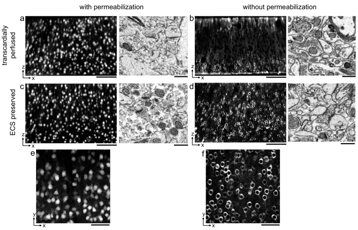
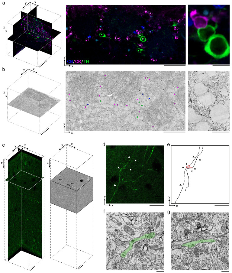
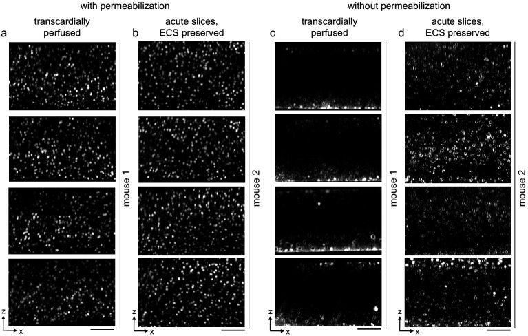
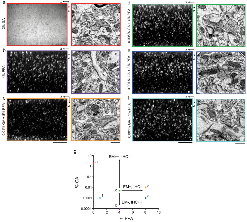
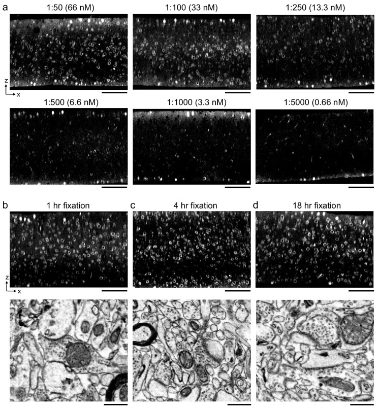
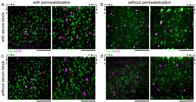
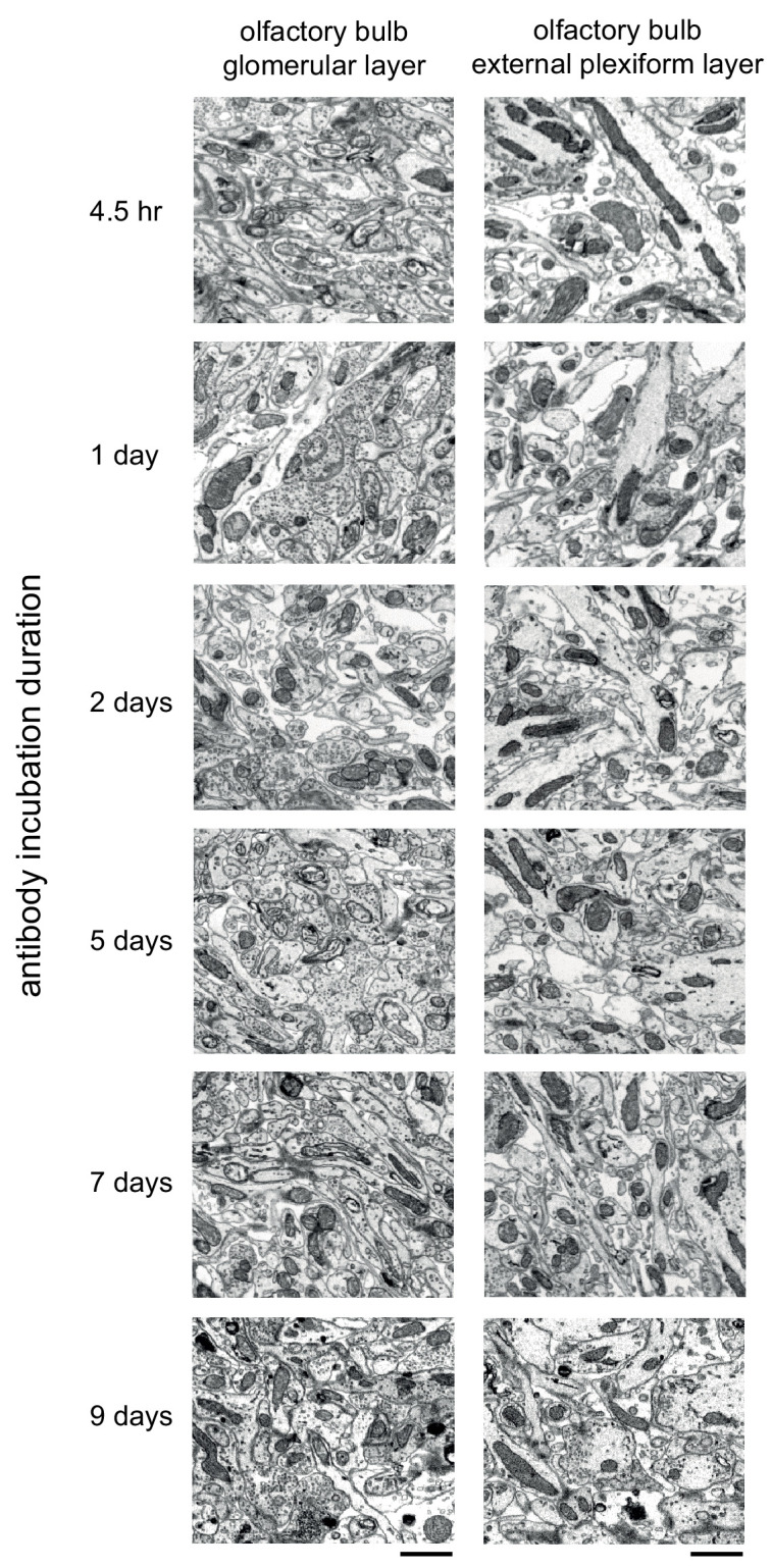
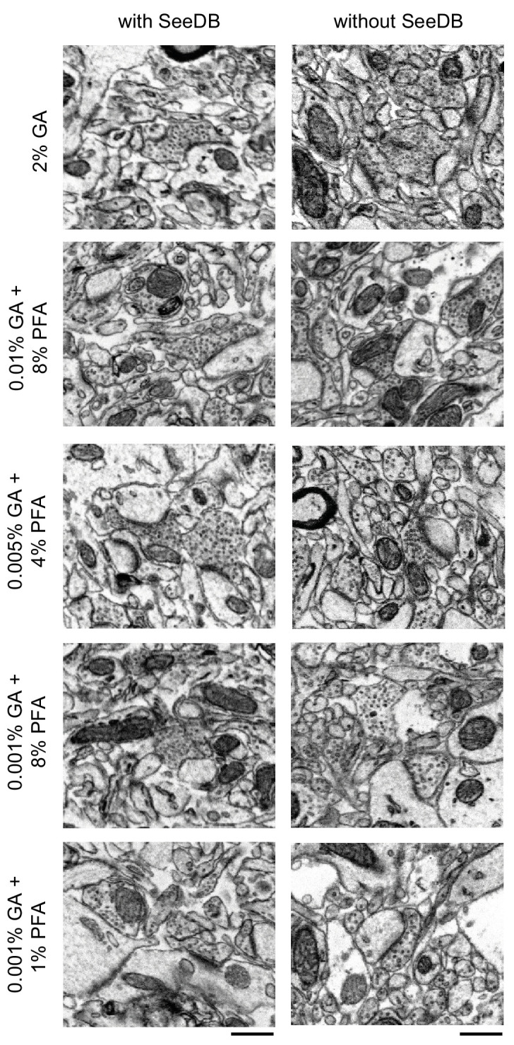
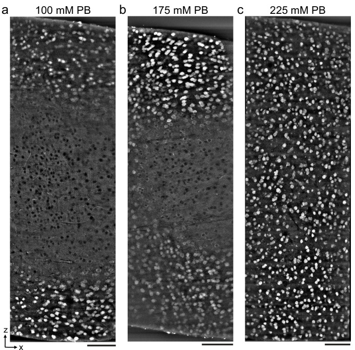
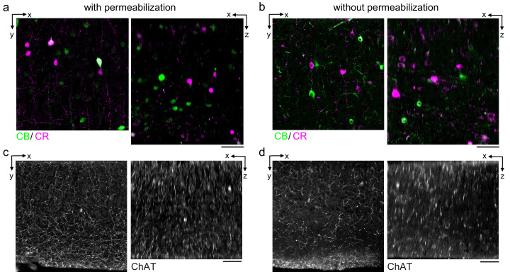
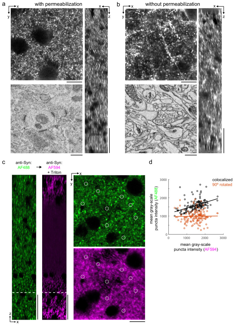
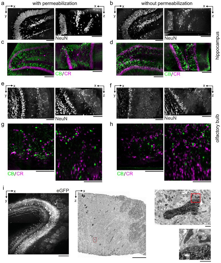
A dense reconstruction of neuronal synaptic connectivity typically requires high-resolution 3D electron microscopy (EM) data, but EM data alone lacks functional information about neurons and synapses. One approach to augment structural EM datasets is with the fluorescent immunohistochemical (IHC) localization of functionally relevant proteins. We describe a protocol that obviates the requirement of tissue permeabilization in thick tissue sections, a major impediment for correlative pre-embedding IHC and EM. We demonstrate the permeabilization-free labeling of neuronal cell types, intracellular enzymes, and synaptic proteins in tissue sections hundreds of microns thick in multiple brain regions from mice while simultaneously retaining the ultrastructural integrity of the tissue. Finally, we explore the utility of this protocol by performing proof-of-principle correlative experiments combining two-photon imaging of protein distributions and 3D EM.
神经元突触连接的密集重建通常需要高分辨率的 3D 电子显微镜 (EM) 数据,但 EM 数据本身缺乏关于神经元和突触的功能信息。一种增强结构 EM 数据集的方法是使用荧光免疫组织化学 (IHC) 对功能相关蛋白进行定位。我们描述了一种方案,该方案避免了在厚组织切片中进行组织通透的要求,这是共定位预包埋 IHC 和 EM 的主要障碍。我们在来自小鼠的多个脑区的数百微米厚的组织切片中演示了神经元细胞类型、细胞内酶和突触蛋白的无通透标记,同时保持组织的超微结构完整性。最后,我们通过进行结合蛋白分布的双光子成像和 3D EM 的原理验证相关性实验来探索该方案的实用性。