Wu Ying-Ying, Chen Chao, Yu Xiao, Zhao Xiao-Dong, Bao Rong-Qi, Yu Jia-Yu, Zhang Guo-Xing, Chen Jing-Wei
Department of Internal Medicine, The Affiliated Suzhou Chinese Traditional Medicine Hospital, Nanjing University of Chinese Medicine, 18 Yang-Su Road, Suzhou 215003, China.
Laboratory of Cancer Molecular Genetics, Medical College of Soochow University, 199 Ren-Ai Road, Dushu Lake Campus, Suzhou Industrial Park, Suzhou 215123, China.
Evid Based Complement Alternat Med. 2018 Jul 12;2018:1765731. doi: 10.1155/2018/1765731. eCollection 2018.
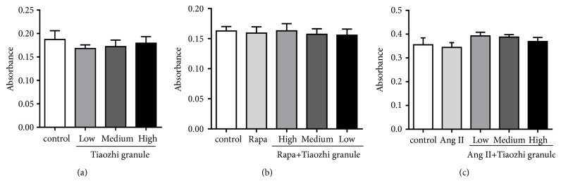
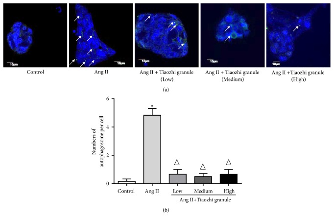
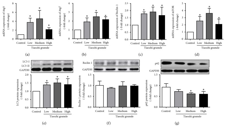
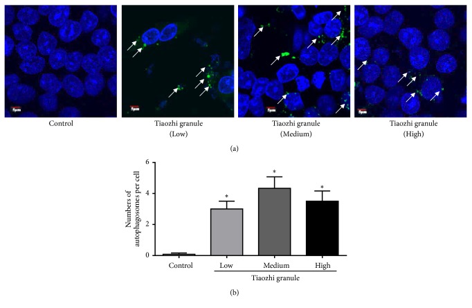
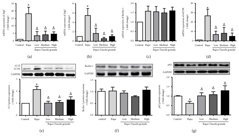
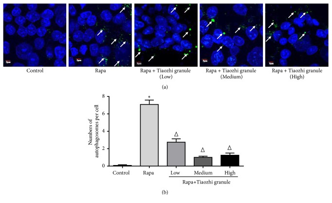
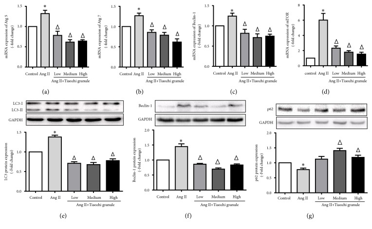
Sera from the rats with Tiaozhi granule treatment were collected. Human umbilical vein endothelial cells (HUVECs) were incubated with different dosage of sera with Tiaozhi granule for 48 hours. Rapamycin or angiotensin II was applied to activate autophagy in HUVECs with or without different dosages of sera of Tiaozhi granule. The mRNA expressions of Atg5, Atg7, Beclin-1, and mammal target of rapamycin (mTOR) were detected by real-time PCR. Autophagic flux markers (protein expression of LC3, Beclin-1, and p62) were examined by western blot analyses. The number of autophagosomes was visualized by immunofluorescence analysis with LC3-II labelling. Results showed that Tiaozhi granule sera increase cell autophagic levels by increase of mRNA of Atg5, Atg7, Beclin-1, and mTOR and increase of autophagic flux and also number of autophagosomes. However, in response to rapamycin or Ang II stimulation, activated autophagic levels were alleviated by Tiaozhi granule sera by reduction of mRNA of Atg5, Atg7, Beclin-1, mTOR, autophagic flux, and also number of autophagosomes. Our present data demonstrate that Tiaozhi granule plays a dual role in response to different cell conditions, which is to increase cell autophagy under physiological condition and to suppress cell excessive autophagy under pathological condition.
收集接受调脂颗粒治疗的大鼠的血清。将人脐静脉内皮细胞(HUVECs)与不同剂量的调脂颗粒血清孵育48小时。应用雷帕霉素或血管紧张素II在有或无不同剂量调脂颗粒血清的情况下激活HUVECs中的自噬。通过实时PCR检测Atg5、Atg7、Beclin-1和雷帕霉素哺乳动物靶蛋白(mTOR)的mRNA表达。通过蛋白质印迹分析检测自噬通量标志物(LC3、Beclin-1和p62的蛋白表达)。通过用LC3-II标记的免疫荧光分析观察自噬体的数量。结果显示,调脂颗粒血清通过增加Atg5、Atg7、Beclin-1和mTOR的mRNA以及增加自噬通量和自噬体数量来提高细胞自噬水平。然而,在雷帕霉素或Ang II刺激下,调脂颗粒血清通过降低Atg5、Atg7、Beclin-1、mTOR的mRNA、自噬通量和自噬体数量来减轻激活的自噬水平。我们目前的数据表明,调脂颗粒在不同细胞条件下发挥双重作用,即在生理条件下增加细胞自噬,在病理条件下抑制细胞过度自噬。