Gu Yue, Ma Lian-Jun, Bai Xiao-Xue, Jie Jing, Zhang Xiu-Fang, Chen Dong, Li Xiao-Ping
Department of Respiratory Medicine, the First Hospital of Jilin University, Changchun, Jilin Province, China.
Endoscopy Center, the China-Japan Hospital of Jilin University, Changchun, Jilin Province, China.
Neural Regen Res. 2018 Oct;13(10):1842-1850. doi: 10.4103/1673-5374.238621.
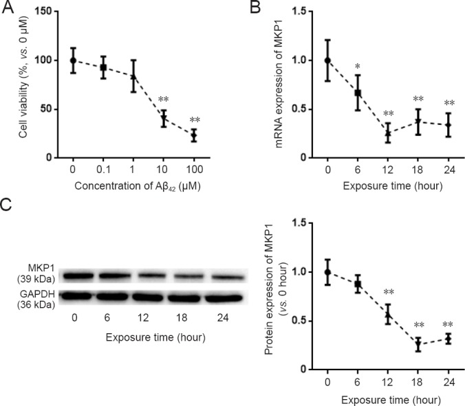
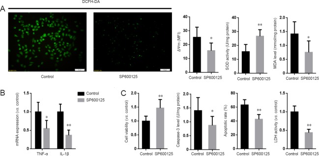
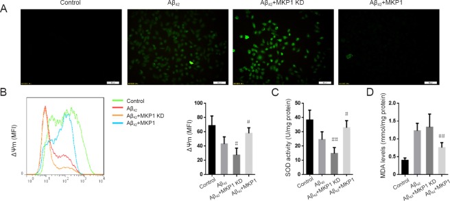
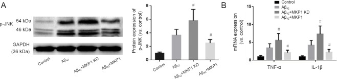
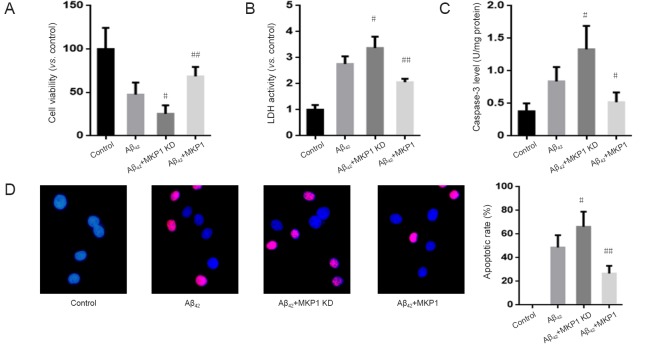
The mitogen-activated protein kinase (MAPK) signaling pathway plays an important role in the regulation of cell growth, proliferation, differentiation, transformation and death. Mitogen-activated protein kinase phosphatase 1 (MKP1) has an inhibitory effect on the p38MAPK and JNK pathways, but it is unknown whether it plays a role in Aβ-induced oxidative stress and neuronal inflammation. In this study, PC12 cells were infected with MKP1 shRNA, MKP1 lentivirus or control lentivirus for 12 hours, and then treated with 0.1, 1, 10 or 100 μM amyloid beta 42 (Aβ). The cell survival rate was measured using the cell counting kit-8 assay. MKP1, tumor necrosis factor-alpha (TNF-α) and interleukin-1β (IL-1β) mRNA expression levels were analyzed using quantitative real time-polymerase chain reaction. MKP1 and phospho-c-Jun N-terminal kinase (JNK) expression levels were assessed using western blot assay. Reactive oxygen species (ROS) levels were detected using 2',7'-dichlorofluorescein diacetate. Mitochondrial membrane potential was measured using flow cytometry. Superoxide dismutase activity and malondialdehyde levels were evaluated using the colorimetric method. Lactate dehydrogenase activity was measured using a microplate reader. Caspase-3 expression levels were assessed by enzyme-linked immunosorbent assay. Apoptosis was evaluated using the terminal deoxynucleotidyl transferase dUTP nick end labeling method. MKP1 overexpression inhibited Aβ-induced JNK phosphorylation and the increase in ROS levels. It also suppressed the Aβ-induced increase in TNF-α and IL-1β levels as well as apoptosis in PC12 cells. In contrast, MKP1 knockdown by RNA interference aggravated Aβ-induced oxidative stress, inflammation and cell damage in PC12 cells. Furthermore, the JNK-specific inhibitor SP600125 abolished this effect of MKP1 knockdown on Aβ-induced neurotoxicity. Collectively, these results show that MKP1 mitigates Aβ-induced apoptosis, oxidative stress and neuroinflammation by inhibiting the JNK signaling pathway, thereby playing a neuroprotective role.
丝裂原活化蛋白激酶(MAPK)信号通路在细胞生长、增殖、分化、转化及死亡的调控中发挥着重要作用。丝裂原活化蛋白激酶磷酸酶1(MKP1)对p38MAPK和JNK信号通路具有抑制作用,但MKP1是否在β淀粉样蛋白(Aβ)诱导的氧化应激及神经元炎症中发挥作用尚不清楚。在本研究中,PC12细胞用MKP1短发夹RNA(shRNA)、MKP1慢病毒或对照慢病毒感染12小时,然后用0.1、1、10或100μM的淀粉样β蛋白42(Aβ)进行处理。使用细胞计数试剂盒-8检测法测定细胞存活率。采用定量实时聚合酶链反应分析MKP1、肿瘤坏死因子-α(TNF-α)和白细胞介素-1β(IL-1β)的mRNA表达水平。用蛋白质免疫印迹法评估MKP1和磷酸化c-Jun氨基末端激酶(JNK)的表达水平。使用2',7'-二氯荧光素二乙酸酯检测活性氧(ROS)水平。采用流式细胞术测量线粒体膜电位。用比色法评估超氧化物歧化酶活性和丙二醛水平。使用酶标仪测量乳酸脱氢酶活性。通过酶联免疫吸附测定法评估半胱天冬酶-3的表达水平。采用末端脱氧核苷酸转移酶dUTP缺口末端标记法评估细胞凋亡。MKP1过表达抑制了Aβ诱导的JNK磷酸化及ROS水平的升高。它还抑制了Aβ诱导的PC12细胞中TNF-α和IL-1β水平的升高以及细胞凋亡。相反,RNA干扰敲低MKP1加重了Aβ诱导的PC12细胞氧化应激、炎症及细胞损伤。此外,JNK特异性抑制剂SP600125消除了MKP1敲低对Aβ诱导的神经毒性的这种作用。总的来说,这些结果表明MKP1通过抑制JNK信号通路减轻Aβ诱导的细胞凋亡、氧化应激及神经炎症,从而发挥神经保护作用。