Istituto Italiano di Tecnologia, Via Morego 30, 16163, Genova, Italy.
INCLIVA Instituto de Investigación Sanitaria, Av. Menéndez Pelayo 4, 46010, Valencia, Spain.
Sci Rep. 2018 Aug 23;8(1):12652. doi: 10.1038/s41598-018-31165-3.
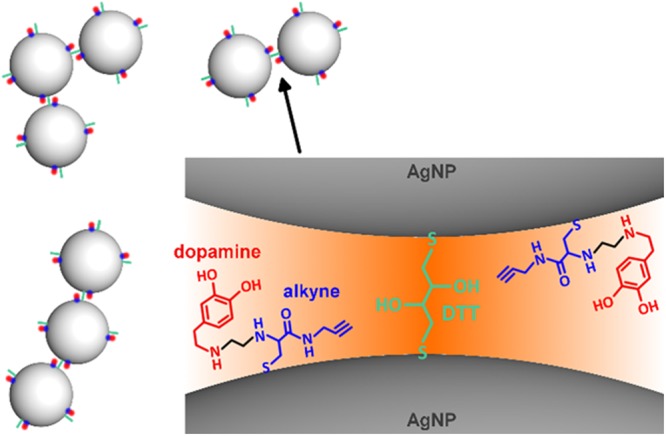
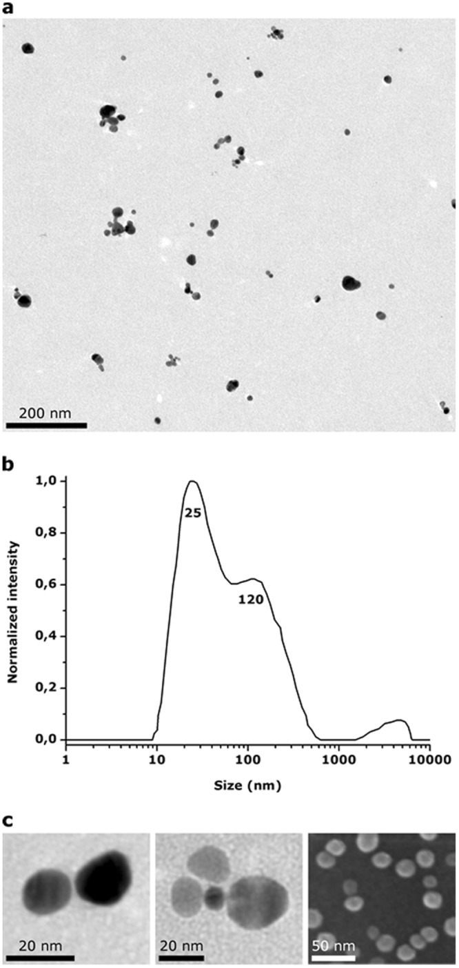
Live intracellular imaging is a valuable tool in modern diagnostics and pharmacology. Surface Enhanced Raman Spectroscopy (SERS) stands out as a non-destructive and multiplexed technique, but intracellular SERS imaging still suffers from interfering background from endogenous components. Here we show the assembly of small colloidal SERS probes with Raman signal in the cell-silent window of 1800-2900 cm for biorthogonal intracellular SERS imaging of dopamine that was undistinguishable from the endogenous cell background. By linking colloidal silver nanoparticles with alkyne-dopamine adducts, clusters are formed by 2-6 nanoparticles spaced by tight interparticle gaps that exhibited high electric field enhancement and strong SERS signals of alkyne and dopamines. Due to the cell-silent signals of the alkyne, intracellular in-vitro Raman imaging shows that the dopamines on the internalized clusters remain distinguishable across the cytoplasm with good spatial resolution. Our method can be a general-purpose method for real-time imaging of biomolecules, such as proteins, peptides, DNA and drugs.
活细胞内成像技术是现代诊断学和药理学中的一种重要工具。表面增强拉曼光谱(SERS)是一种非破坏性和多重的技术,但细胞内 SERS 成像仍然受到内源性成分干扰背景的影响。在这里,我们展示了具有在细胞静默窗口 1800-2900cm 处拉曼信号的小胶体 SERS 探针的组装,用于多巴胺的生物正交细胞内 SERS 成像,其与内源性细胞背景无法区分。通过将银纳米颗粒与炔基-多巴胺加合物连接,由 2-6 个纳米颗粒组成的簇通过紧密的颗粒间间隙隔开,表现出高电场增强和炔基和多巴胺的强 SERS 信号。由于炔基的细胞静默信号,细胞内的体外拉曼成像表明,内化簇上的多巴胺在细胞质中仍然可以通过良好的空间分辨率来区分。我们的方法可以成为实时成像生物分子(如蛋白质、肽、DNA 和药物)的通用方法。