Kwak Min-Sun, Chung Goh Eun, Chung Su Jin, Kang Seung Joo, Yang Jong In, Kim Joo Sung
Department of Internal Medicine, Healthcare Research Institute, Healthcare System Gangnam Center, Seoul National University Hospital, Seoul, Republic of Korea.
Gastroenterol Res Pract. 2018 Jul 24;2018:8796165. doi: 10.1155/2018/8796165. eCollection 2018.
and gastric atrophy are risk factors for gastric cancer. We evaluated whether the combination of serum HP antibody and pepsinogen (PG), which is indicative of gastric atrophy, could serve as a predictive marker for the development of gastric neoplasms in a Korean population.
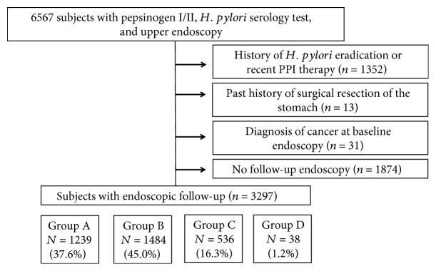
The subjects who had undergone health-screening examination with endoscopic follow-ups were classified into the following 4 groups according to serum PG status and antibody at baseline: group A ( (-), normal PG), group B ( (+), normal PG), group C ( (+), atrophic PG), and group D ( (-), atrophic PG). We compared the development of gastric neoplasms among the groups.
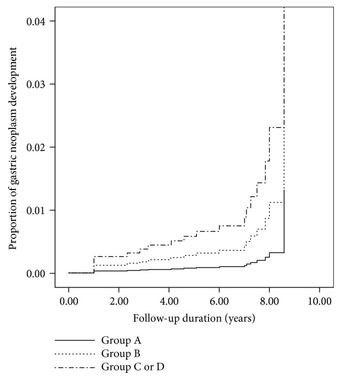
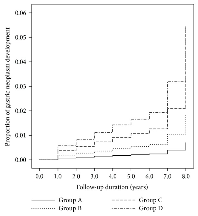
Of the 3297 subjects, 1239 (37.6%) were categorized as group A, 1484 (45.0%) as group B, 536 (16.3%) as group C, and 38 (1.2%) as group D. During the 5.6 years of mean follow-up period, the annual incidence of gastric neoplasms increased gradually by 0.06% in group A, 0.16% in group B, 0.38% in group C, and 0.49% in group D. A Cox proportional hazard model showed increased development of gastric neoplasms according to group ( for trend = 0.025). Compared to group A, the hazard ratio was 8.25 for group D (95% confidence interval 0.2-74.24), 5.35 for group C (1.68-17.05), and 2.65 for group B (0.86-8.14).
The combination of serum PG and antibody is useful for predicting the development of gastric neoplasms, including cancer and adenoma, in a Korean population using endoscopic surveillance.
幽门螺杆菌(HP)感染和胃萎缩是胃癌的危险因素。我们评估了血清HP抗体与提示胃萎缩的胃蛋白酶原(PG)的联合检测能否作为韩国人群胃肿瘤发生的预测标志物。
对接受过内镜随访的健康筛查受试者,根据基线时血清PG状态和HP抗体分为以下4组:A组(HP抗体阴性,PG正常),B组(HP抗体阳性,PG正常),C组(HP抗体阳性,PG萎缩),D组(HP抗体阴性,PG萎缩)。我们比较了各组胃肿瘤的发生情况。
在3297名受试者中,1239人(37.6%)被归类为A组,1484人(45.0%)为B组,536人(16.3%)为C组,38人(1.2%)为D组。在平均5.6年的随访期内,胃肿瘤的年发病率在A组逐渐增加0.06%,B组增加0.16%,C组增加0.38%,D组增加0.49%。Cox比例风险模型显示,胃肿瘤的发生根据分组增加(趋势检验P = 0.025)。与A组相比,D组的风险比为8.25(95%置信区间0.2 - 74.24),C组为5.35(1.68 - 17.05),B组为2.65(0.86 - 8.14)。
血清PG和HP抗体的联合检测有助于通过内镜监测预测韩国人群胃肿瘤(包括癌症和腺瘤)的发生。