Zhang Jing, Fan Yimeng, Song Yanlin, Xu Jianguo
Department of Neurosurgery West China School of Medicine, West China Hospital, Sichuan University, Sichuan, PR China.
Medicine (Baltimore). 2018 Aug;97(34):e12022. doi: 10.1097/MD.0000000000012022.
Raman spectroscopy could be applied to distinguish tumor from normal tissues. This meta-analysis assessed the accuracy of Raman spectroscopy in differentiating skin cancer from normal tissue.
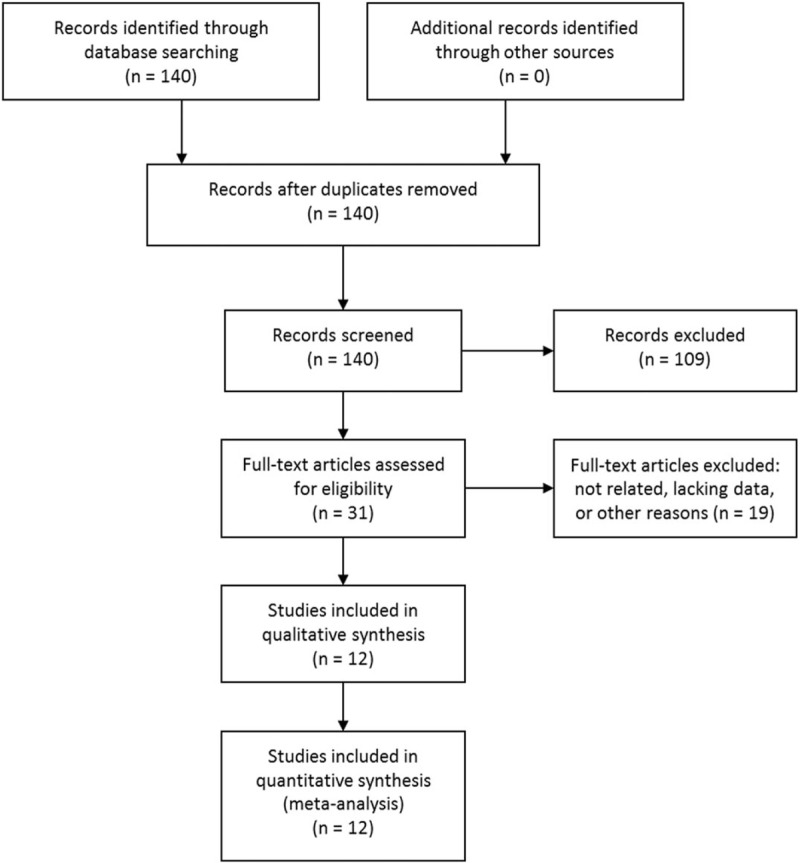
PubMed, Embase, Cochrane Library, and CNKI were searched to identify suitable studies before Februray 4th, 2018. We estimated the pooled sensitivity, specificity, positive, and negative likelihood ratios, diagnostic odds ratio, and constructed summary receiver-operating characteristics curves to identify the accuracy of Raman spectroscopy in differentiating skin cancer from normal tissue.
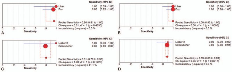
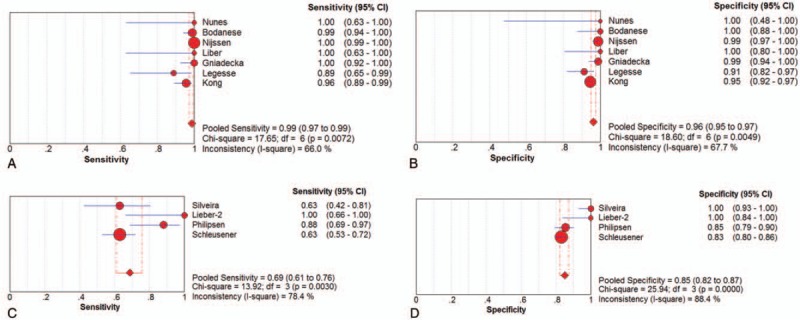
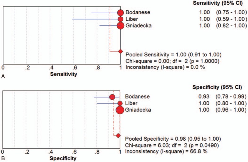
A total of 12 studies with 2461 spectra were included. For basal cell skin cancer (BCC) ex vivo detection, the pooled sensitivity and specificity were 0.99 (95% confidence interval [CI] 0.97-0.99) and 0.96 (95% CI 0.95-0.97), respectively. The area under the curve (AUC) was 0.9837. For BCC in vivo detection, the pooled sensitivity and specificity were 0.69 (95% CI 0.61-0.76) and 0.85 (95% CI 0.82-0.87), respectively. The AUC was 0.9213. For melanoma (MM) ex vivo detection, the pooled sensitivity and specificity were 1.00 (95% CI 0.91-1.00) and 0.98 (95% CI 0.95-1.00), respectively. The AUC was 0.9914. For MM in vivo detection, the sensitivity (0.93) and the specificity (0.96) balanced relatively well. For squamous cell skin cancer (SCC) ex vivo detection, the pooled sensitivity and specificity were 0.96 (95% CI 0.81-1.00) and 1.00 (95% CI 0.92-1.00), respectively. For SCC in vivo detection, the sensitivity was 0.81 (95% CI 0.70-0.90) and the specificity was 0.89 (95% CI 0.86-0.91).
This meta-analysis suggested that Raman spectroscopy could be an effective and accurate tool for differentiating BCC, MM, SCC from normal tissue, which would assist us in the diagnosis and treatment of skin cancer.
拉曼光谱可用于区分肿瘤组织与正常组织。本荟萃分析评估了拉曼光谱在鉴别皮肤癌与正常组织方面的准确性。
检索了PubMed、Embase、Cochrane图书馆和中国知网,以识别2018年2月4日前的合适研究。我们估计了合并敏感性、特异性、阳性和阴性似然比、诊断比值比,并构建了汇总的受试者工作特征曲线,以确定拉曼光谱在鉴别皮肤癌与正常组织方面的准确性。
共纳入12项研究,包含2461个光谱。对于离体检测基底细胞皮肤癌(BCC),合并敏感性和特异性分别为0.99(95%置信区间[CI]0.97 - 0.99)和0.96(95%CI 0.95 - 0.97)。曲线下面积(AUC)为0.9837。对于活体检测BCC,合并敏感性和特异性分别为0.69(95%CI 0.61 - 0.76)和0.85(95%CI 0.82 - 0.87)。AUC为0.9213。对于离体检测黑色素瘤(MM),合并敏感性和特异性分别为1.00(95%CI 0.91 - 1.00)和0.98(95%CI 0.95 - 1.00)。AUC为0.9914。对于活体检测MM,敏感性(0.93)和特异性(0.96)平衡相对较好。对于离体检测鳞状细胞皮肤癌(SCC),合并敏感性和特异性分别为0.96(95%CI 0.81 - 1.00)和1.00(95%CI 0.92 - 1.00)。对于活体检测SCC,敏感性为0.81(95%CI 0.70 - 0.90),特异性为0.89(95%CI 0.86 - 0.91)。
本荟萃分析表明,拉曼光谱可能是一种有效且准确的工具,用于区分BCC、MM、SCC与正常组织,这将有助于我们对皮肤癌的诊断和治疗。