Laboratory of Structural Dynamics, Stability and Folding of Proteins, Institute of Cytology of the Russian Academy of Science, Tikhoretsky ave. 4, 194064 St. Petersburg, Russia.
Institute of Physics, Nanotechnology and Telecommunications, Peter the Great St.-Petersburg Polytechnic University, Polytechnicheskaya 29, 195251 St. Petersburg, Russia.
Int J Mol Sci. 2018 Aug 23;19(9):2486. doi: 10.3390/ijms19092486.
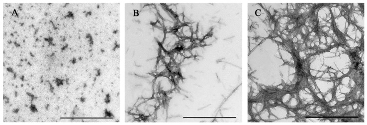
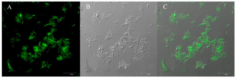
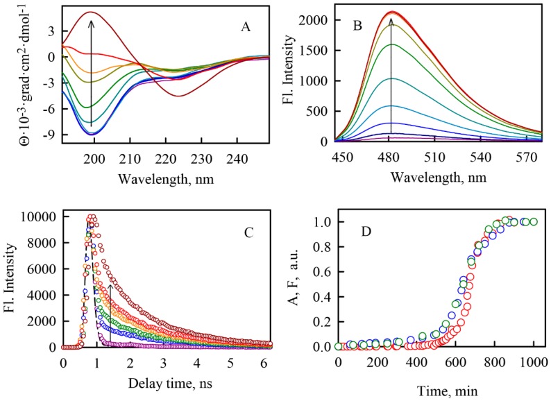
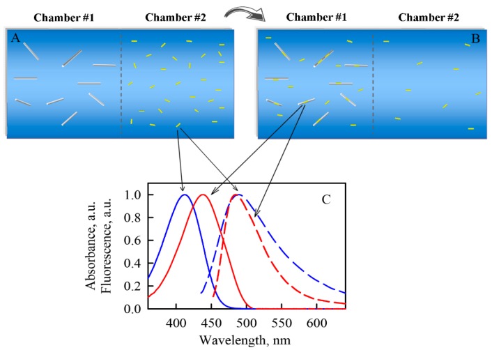
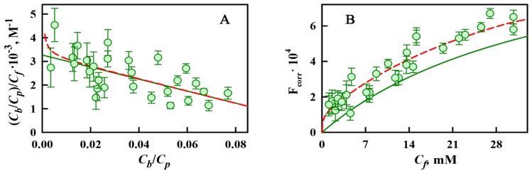
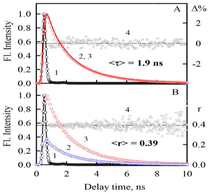
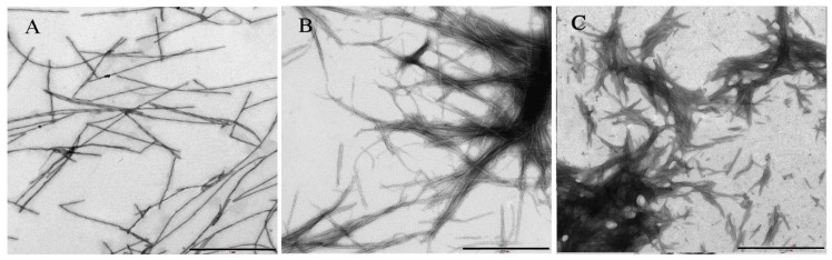
In this work, α-synuclein amyloid fibrils-the formation of which is a biomarker of Parkinson's disease-were investigated using the fluorescent probe thioflavin T (ThT). The experimental conditions of protein fibrillogenesis were chosen so that a sufficient number of continuous measurements could be performed to characterize and analyze all stages of this process. The reproducibility of fibrillogenesis and the structure of the obtained aggregates (which is a critical point for further investigation) were proven using a wide range of physical-chemical methods. For the determination of ThT-α-synuclein amyloid fibril binding parameters, the sample and reference solutions were prepared using equilibrium microdialysis. By utilizing absorption spectroscopy of these solutions, the ThT-fibrils binding mode with a binding constant of about 10⁴ M and stoichiometry of ThT per protein molecule of about 1:8 was observed. Fluorescence spectroscopy of the same solutions with the subsequent correction of the recorded fluorescence intensity on the primary inner filter effect allowed us to determine another mode of ThT binding to fibrils, with a binding constant of about 10⁶ M and stoichiometry of about 1:2500. Analysis of the photophysical characteristics of the dye molecules bound to the sites of different binding modes allowed us to assume the possible localization of these sites. The obtained differences in the ThT binding parameters to the amyloid fibrils formed from α-synuclein and other amyloidogenic proteins, as well as in the photophysical characteristics of the bound dye, confirmed the hypothesis of amyloid fibril polymorphism.
在这项工作中,使用荧光探针硫黄素 T(ThT)研究了α-突触核蛋白淀粉样纤维 - 其形成是帕金森病的生物标志物。选择蛋白质纤维形成的实验条件,以便可以进行足够数量的连续测量,以表征和分析该过程的所有阶段。使用广泛的物理化学方法证明了纤维发生的重现性和获得的聚集体的结构(这是进一步研究的关键点)。使用平衡微透析法制备样品和参比溶液,以确定 ThT-α-突触核蛋白淀粉样纤维结合参数。通过这些溶液的吸收光谱,观察到结合常数约为 10⁴ M,每个蛋白质分子的 ThT 配位数约为 1:8 的 ThT-纤维结合模式。对相同溶液的荧光光谱进行校正后,记录的荧光强度的原始内滤效应,使我们能够确定另一种 ThT 与纤维结合的模式,结合常数约为 10⁶ M,配位数约为 1:2500。分析结合到不同结合模式的染料分子的光物理特性,使我们能够假设这些结合部位的可能定位。从α-突触核蛋白和其他淀粉样蛋白形成的淀粉样纤维中获得的 ThT 结合参数以及结合染料的光物理特性的差异,证实了淀粉样纤维多态性的假说。