Department of Anatomy, Korea University College of Medicine, Seoul, 02841, Republic of Korea.
Department of Physics, Korea University, Seoul, 02841, Republic of Korea.
Sci Rep. 2018 Aug 24;8(1):12815. doi: 10.1038/s41598-018-31153-7.
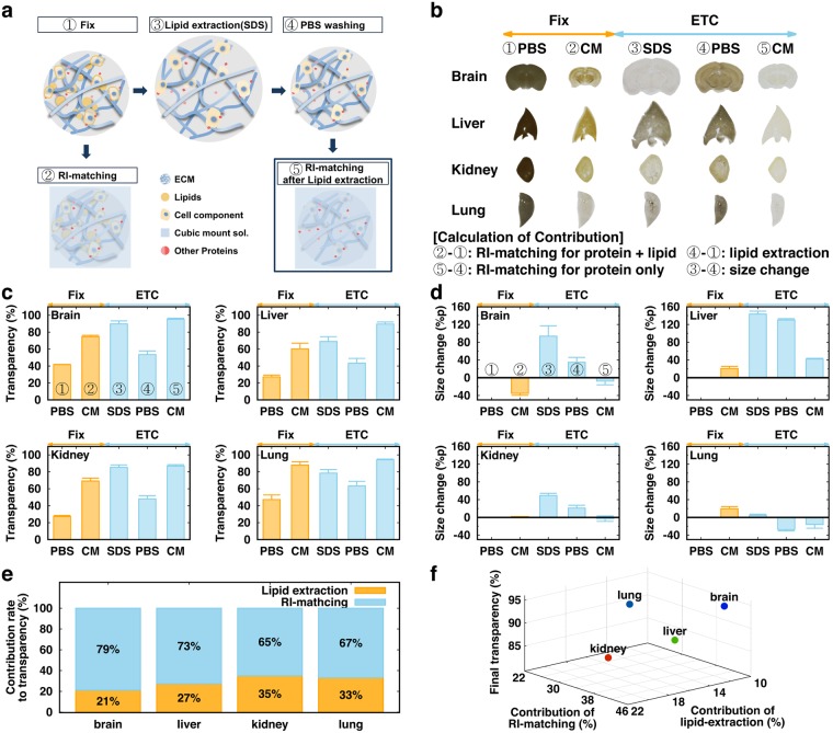
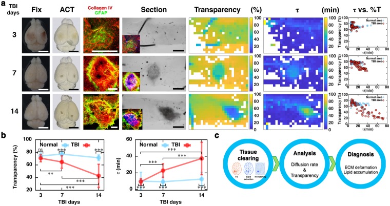
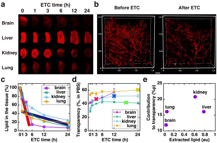
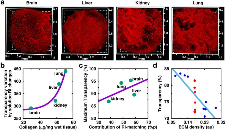
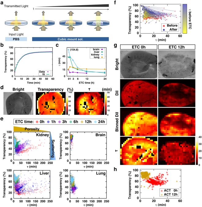
Tissue-clearing techniques have received great attention for volume imaging and for the potential to be applied in optical diagnosis. In principle, tissue clearing is achieved by reducing light scattering through a combination of lipid removal, size change, and matching of the refractive index (RI) between the imaging solution and the tissue. However, the contributions of these major factors in tissue clearing have not been systematically evaluated yet. In this study, we experimentally measured and mathematically calculated the contribution of these factors to the clearing of four organs (brain, liver, kidney, and lung). We found that these factors differentially influence the maximal clearing efficacy of tissues and the diffusivity of materials inside the tissue. We propose that these physical properties of organs can be utilized for the quality control (Q/C) process during tissue clearing, as well as for the monitoring of the pathological changes of tissues.
组织透明化技术因其在容积成像方面的应用和在光学诊断方面的应用潜力而备受关注。从原理上讲,组织透明化是通过去除脂质、改变大小以及匹配成像溶液和组织之间的折射率 (RI) 来减少光散射来实现的。然而,这些主要因素在组织透明化中的贡献尚未得到系统评估。在这项研究中,我们通过实验测量和数学计算评估了这些因素对四种器官(脑、肝、肾和肺)的清除效果的贡献。我们发现这些因素会对组织的最大清除效果和组织内部物质的扩散性产生不同的影响。我们提出,这些器官的物理特性可以用于组织透明化过程中的质量控制 (Q/C) 以及组织病理变化的监测。