Sepsi Adél, Fábián Attila, Jäger Katalin, Heslop-Harrison J S, Schwarzacher Trude
Department of Plant Cell Biology, Centre for Agricultural Research, Hungarian Academy of Sciences, Martonvásár, Hungary.
Department of Genetics and Genome Biology, University of Leicester, Leicester, United Kingdom.
Front Plant Sci. 2018 Aug 14;9:1193. doi: 10.3389/fpls.2018.01193. eCollection 2018.
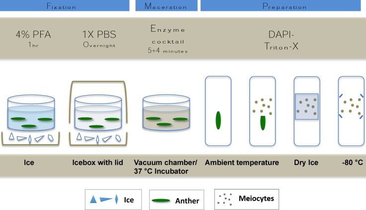
ImmunoFISH is a method combining immunolabelling (IL) with fluorescent hybridisation (FISH) to simultaneously detect the nuclear distribution of proteins and specific DNA sequences within chromosomes. This approach is particularly important when analysing meiotic cell division where morphogenesis of individual proteins follows stage-specific changes and is accompanied by a noticeable chromatin dynamism. The method presented here is simple and provides reliable results of high quality signal, low background staining and can be completed within 2 days following preparation. Conventional widefield epifluorescent or laser scanning microscopy can be used for high resolution and three-dimensional analysis. Fixation and preparation techniques were optimised to best preserve nuclear morphology and protein epitopes without the need for any antigen retrieval. Preparation of plant material involved short cross-linking fixation of meiotic tissues with paraformaldehyde (PFA) followed by enzyme digestion and slide-mounting. In order to avoid rapid sample degradation typical of shortly fixed plant materials, and to be able to perform IL later, slides were snap-frozen and stored at -80°C. Ultra-freezing produced a remarkable degree of structural preservation for up to 12 months, whereby sample quality was similar to that of fresh material. Harsh chemicals and sample dehydration were avoided throughout the procedure and permeability was ensured by a 0.1-0.3% detergent treatment. The ImmunoFISH method was developed specifically for studying meiosis in , but should also be applicable to other grass and plant species.
免疫荧光原位杂交(ImmunoFISH)是一种将免疫标记(IL)与荧光杂交(FISH)相结合的方法,用于同时检测蛋白质的核分布和染色体中的特定DNA序列。在分析减数分裂细胞分裂时,这种方法尤为重要,因为单个蛋白质的形态发生会随阶段特异性变化,并且伴随着明显的染色质动态变化。这里介绍的方法简单,能提供高质量信号、低背景染色的可靠结果,并且在样品制备后2天内即可完成。传统的宽场落射荧光显微镜或激光扫描显微镜可用于高分辨率和三维分析。固定和制备技术经过优化,能在无需任何抗原修复的情况下,最佳地保存核形态和蛋白质表位。植物材料的制备包括用多聚甲醛(PFA)对减数分裂组织进行短时间交联固定,然后进行酶消化和载玻片固定。为了避免短时间固定的植物材料典型的快速样品降解,并能够随后进行免疫标记,载玻片被速冻并储存在-80°C。超低温冷冻可在长达12个月的时间内实现显著程度的结构保存,从而使样品质量与新鲜材料相似。在整个过程中避免使用苛刻的化学试剂和样品脱水,并通过0.1 - 0.3%的去污剂处理确保通透性。免疫荧光原位杂交方法是专门为研究[具体植物名称未给出]的减数分裂而开发的,但也应适用于其他禾本科植物和植物物种。