Guo Yanan, Beyle Franziska E, Bold Beatrix M, Watanabe Hiroshi C, Koslowski Axel, Thiel Walter, Hegemann Peter, Marazzi Marco, Elstner Marcus
Department of Theoretical Chemical Biology , Institute of Physical Chemistry , KIT , Kaiserstrasse 12 , 76131 Karlsruhe , Germany . Email:
Research Center for Advanced Science and Technology , The University of Tokyo , 4-6-1 Komaba, Meguro-ku , Tokyo 153-8904 , Japan.
Chem Sci. 2016 Jun 1;7(6):3879-3891. doi: 10.1039/c6sc00468g. Epub 2016 Feb 26.
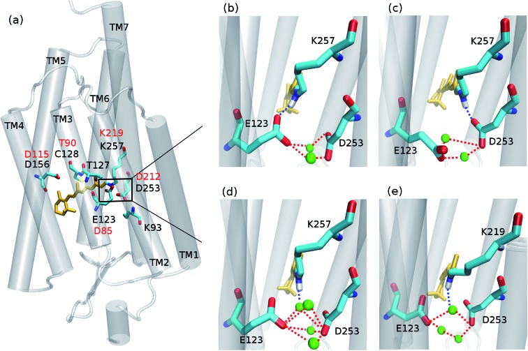
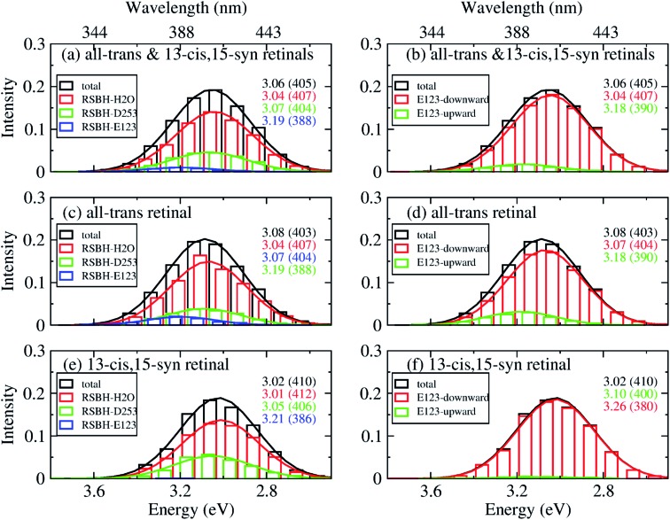
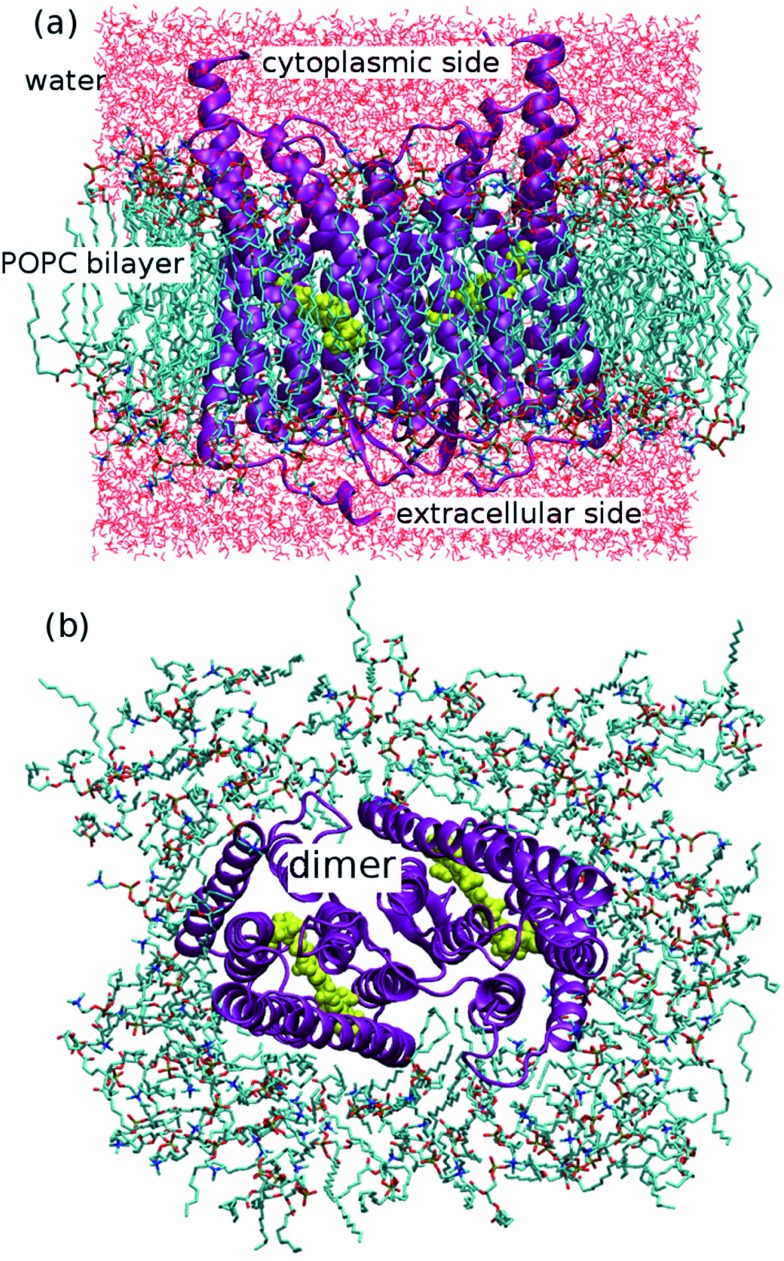
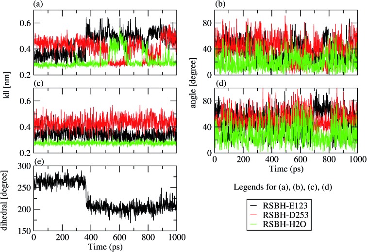
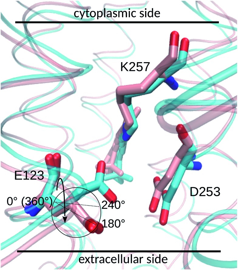
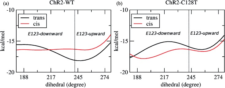
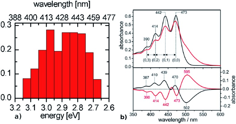
In spite of considerable interest, the active site of channelrhodopsin still lacks a detailed atomistic description, the understanding of which could strongly enhance the development of novel optogenetics tools. We present a computational study combining different state-of-the-art techniques, including hybrid quantum mechanics/molecular mechanics schemes and high-level quantum chemical methods, to properly describe the hydrogen-bonding pattern between the retinal chromophore and its counterions in channelrhodopsin-2 Wild-Type and C128T mutant. Especially, we show by extensive ground state dynamics that the active site, containing a glutamic acid (E123) and a water molecule, is highly dynamic, sampling three different hydrogen-bonding patterns. This results in a broad absorption spectrum that is representative of the different structural motifs found. A comparison with bacteriorhodopsin, characterized by a pentagonal hydrogen-bonded active site structure, elucidates their different absorption properties.
尽管备受关注,但通道视紫红质的活性位点仍缺乏详细的原子水平描述,对其的理解可能会极大地促进新型光遗传学工具的开发。我们开展了一项计算研究,结合了不同的前沿技术,包括混合量子力学/分子力学方案和高级量子化学方法,以准确描述通道视紫红质-2野生型和C128T突变体中视黄醛发色团与其抗衡离子之间的氢键模式。特别是,我们通过广泛的基态动力学表明,包含谷氨酸(E123)和一个水分子的活性位点具有高度动态性,呈现出三种不同的氢键模式。这导致了一个宽泛的吸收光谱,该光谱代表了所发现的不同结构基序。与以五角形氢键活性位点结构为特征的细菌视紫红质进行比较,阐明了它们不同的吸收特性。