Department of Biology, American University, 4400 Massachusetts Ave NW, Washington, DC 20016, USA.
Neural Circuitry Unit, National Institute of Neurological Disorders and Stroke, National Institutes of Health, 5625 Fisher's Lane, Rockville, MD 20852, USA.
Dis Model Mech. 2018 Oct 22;11(10):dmm035220. doi: 10.1242/dmm.035220.
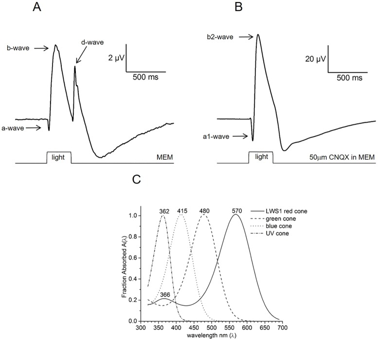
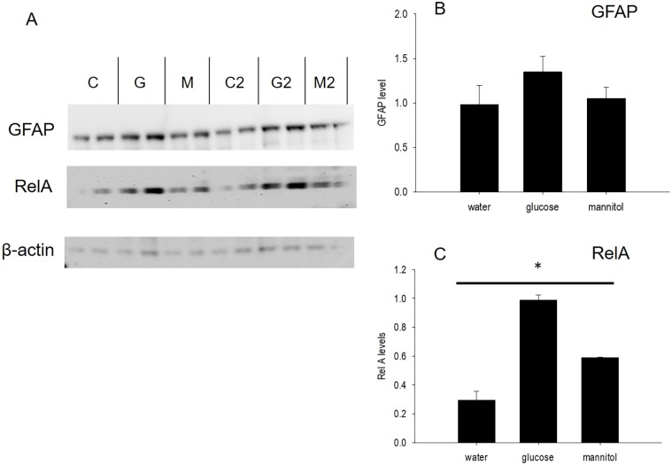
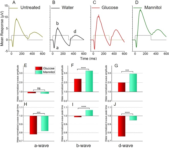
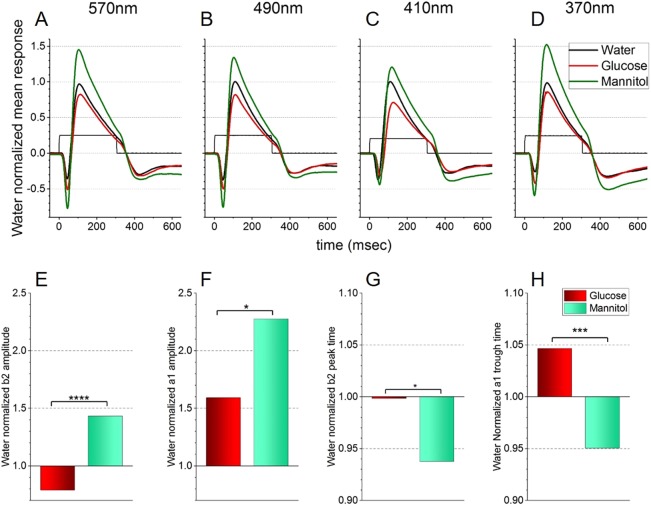
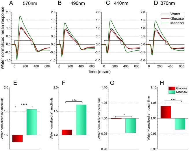
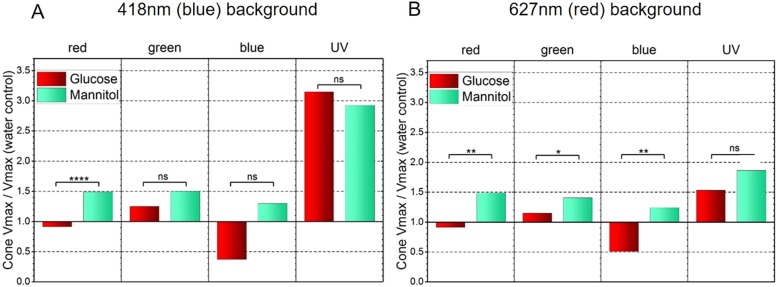
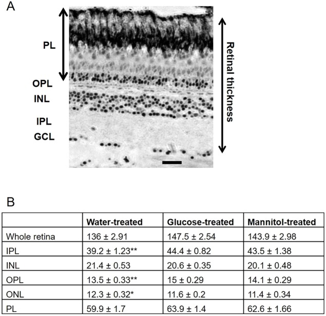
Prolonged hyperglycemia can alter retinal function, ultimately resulting in blindness. Adult zebrafish adults exposed to alternating conditions of 2% glucose/0% glucose display a 3× increase in blood sugar levels. After 4 weeks of treatment, electroretinograms (ERGs) were recorded from isolated, perfused, eyecups. Control animals were exposed to alternating 2% mannitol/0% mannitol (osmotic control) or to alternating water (0% glucose/0% glucose; handling control). Two types of ERGs were recorded: (1) native ERGs measured using white-light stimuli and medium without synaptic blockers; and (2) spectral ERGs measured with an AMPA/kainate receptor antagonist, isolating photoreceptor-to-ON-bipolar-cell synapses, and a spectral protocol that separated red (R), green (G), blue (B) and UV cone signals. Retinas were evaluated for changes in layer thickness and for the inflammatory markers GFAP and Nf-κB (RelA or p65). In native ERGs, hyperglycemic b- and d-waves were lower in amplitude than the b- and d-waves of mannitol controls. Alteration of waveshape became severe, with b-waves becoming more transient and ERG responses showing more PIII-like (a-wave) characteristics. For spectral ERGs, waveshape appeared similar in all treatment groups. However, a1- and b2-wave implicit times were significantly longer, and amplitudes were significantly reduced, in response to hyperglycemic treatment, owing to the functional reduction in signals from R, G and B cones. Nf-κB increased significantly in hyperglycemic retinas, but the increase in GFAP was not significant and retinal layer thickness was unaffected. Thus, prolonged hyperglycemia triggers an inflammatory response and functional deficits localized to specific cone types, indicating the rapid onset of neural complications in the zebrafish model of diabetic retinopathy.
长期的高血糖会改变视网膜功能,最终导致失明。成年斑马鱼在 2%葡萄糖/0%葡萄糖的交替条件下暴露,血糖水平会增加 3 倍。经过 4 周的治疗,从分离的灌注眼杯中记录视网膜电图(ERG)。对照动物分别暴露于 2%甘露醇/0%甘露醇(渗透对照)或水(0%葡萄糖/0%葡萄糖;处理对照)的交替环境中。记录了两种类型的 ERG:(1)使用白光刺激和无突触阻滞剂的介质测量的原生 ERG;(2)用 AMPA/kainate 受体拮抗剂测量的光谱 ERG,分离光感受器到 ON-双极细胞突触,并使用分离红(R)、绿(G)、蓝(B)和紫外锥信号的光谱方案。评估视网膜厚度变化以及炎症标志物 GFAP 和 Nf-κB(RelA 或 p65)。在原生 ERG 中,高血糖 b-和 d-波的幅度低于甘露醇对照的 b-和 d-波。波型变化变得严重,b-波变得更短暂,ERG 反应显示出更多的 PIII 样(a-波)特征。对于光谱 ERG,在所有治疗组中波型似乎相似。然而,由于 R、G 和 B 锥信号的功能降低,a1-和 b2-波的潜伏期显著延长,振幅显著降低。在高血糖处理下,Nf-κB 显著增加,但 GFAP 的增加不显著,视网膜层厚度不受影响。因此,长期高血糖会引发炎症反应和特定锥体细胞类型的功能缺陷,表明糖尿病性视网膜病变斑马鱼模型中神经并发症的快速发生。