Department of Neuroradiology, Heidelberg University Hospital, Im Neuenheimer Feld 400, 69120, Heidelberg, Germany.
Division of Experimental Radiology, Department of Neuroradiology, Heidelberg University Hospital, Im Neuenheimer Feld 400, 69120, Heidelberg, Germany.
Sci Rep. 2018 Aug 29;8(1):13029. doi: 10.1038/s41598-018-31384-8.
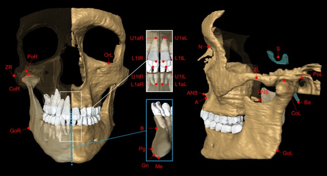
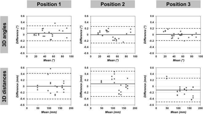
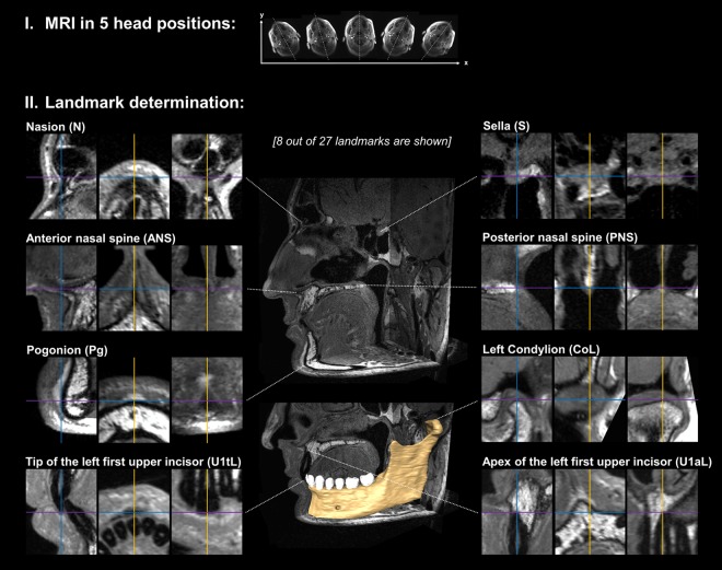
The aim of this study was to validate geometric accuracy and in vivo reproducibility of landmark-based cephalometric measurements using high-resolution 3D Magnetic Resonance Imaging (MRI) at 3 Tesla. For accuracy validation, 96 angular and 96 linear measurements were taken on a phantom in 3 different positions. In vivo MRI scans were performed on 3 volunteers in five head positions. For each in vivo scan, 27 landmarks were determined from which 19 angles and 26 distances were calculated. Statistical analysis was performed using Bland-Altman analysis, the two one-sided tests procedure and repeated measures one-way analysis of variance. In comparison to ground truth, all MRI-based phantom measurements showed statistical equivalence (p < 0.001) and an excellent agreement in Bland-Altman analysis (bias ranges: -0.090-0.044°, -0.220-0.241 mm). In vivo cephalometric analysis was highly reproducible among the five different head positions in all study participants, without statistical differences for all angles and distances (p > 0.05). Ranges between maximum and minimum in vivo values were consistently smaller than 2° and 2 mm, respectively (average ranges: 0.88°/0.87 mm). In conclusion, this study demonstrates that accurate and reproducible 3D cephalometric analysis can be performed without exposure to ionizing radiation using MRI.
本研究旨在验证基于标志点的头影测量在 3T 高分辨率 3D 磁共振成像(MRI)中的几何精度和体内可重复性。为了进行准确性验证,在 3 个不同位置的 96 个角测量和 96 个线测量中进行了 96 个角测量和 96 个线测量。在 3 名志愿者的 5 个头位进行了体内 MRI 扫描。对于每个体内扫描,从 27 个标志点确定了 19 个角度和 26 个距离。使用 Bland-Altman 分析、双单边检验程序和重复测量单向方差分析进行统计分析。与真实值相比,所有基于 MRI 的体模测量均显示出统计学等效性(p<0.001),Bland-Altman 分析也具有极好的一致性(偏差范围:-0.090-0.044°,-0.220-0.241 mm)。在所有研究参与者的 5 个不同头位中,体内头影测量分析具有高度可重复性,所有角度和距离均无统计学差异(p>0.05)。体内测量的最大和最小范围始终小于 2°和 2 mm(平均范围:0.88°/0.87 mm)。总之,本研究表明,使用 MRI 可以在不暴露于电离辐射的情况下进行准确且可重复的 3D 头影测量分析。