• 文献检索
  • 文档翻译
  • 深度研究
  • 学术资讯
  • Suppr Zotero 插件Zotero 插件
  • 邀请有礼
  • 套餐&价格
  • 历史记录
应用&插件
Suppr Zotero 插件Zotero 插件浏览器插件Mac 客户端Windows 客户端微信小程序
定价
高级版会员购买积分包购买API积分包
服务
文献检索文档翻译深度研究API 文档MCP 服务
关于我们
关于 Suppr公司介绍联系我们用户协议隐私条款
关注我们

Suppr 超能文献

核心技术专利:CN118964589B侵权必究
粤ICP备2023148730 号-1Suppr @ 2026

文献检索

告别复杂PubMed语法,用中文像聊天一样搜索,搜遍4000万医学文献。AI智能推荐,让科研检索更轻松。

立即免费搜索

文件翻译

保留排版,准确专业,支持PDF/Word/PPT等文件格式,支持 12+语言互译。

免费翻译文档

深度研究

AI帮你快速写综述,25分钟生成高质量综述,智能提取关键信息,辅助科研写作。

立即免费体验

眼前节 OCT 下微创经巩膜内路 Glaucoma Gel Microstent 植入术后房水引流物的纵向形态。

Longitudinal bleb morphology in anterior segment OCT after minimally invasive transscleral ab interno Glaucoma Gel Microstent implantation.

机构信息

Department of Ophthalmology and Optometry, University Clinic Salzburg, Paracelsus Medical University, Salzburg, Austria.

Research Program Experimental Ophthalmology and Glaucoma Research, Paracelsus Medical University, Salzburg, Austria.

出版信息

Acta Ophthalmol. 2019 Mar;97(2):e231-e237. doi: 10.1111/aos.13902. Epub 2018 Aug 29.

DOI:10.1111/aos.13902
PMID:30160048
原文链接:https://pmc.ncbi.nlm.nih.gov/articles/PMC6586011/
Abstract

PURPOSE

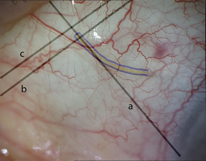
Like the classic trabeculectomy, the minimally invasive, ab interno XEN Glaucoma Gel Microstent (XEN-GGM) creates a filtration bleb in the conjunctiva. The goal of this study was to investigate internal bleb morphology over time with anterior segment optical coherence tomography (AS-OCT) after XEN-GGM implantation.

METHODS

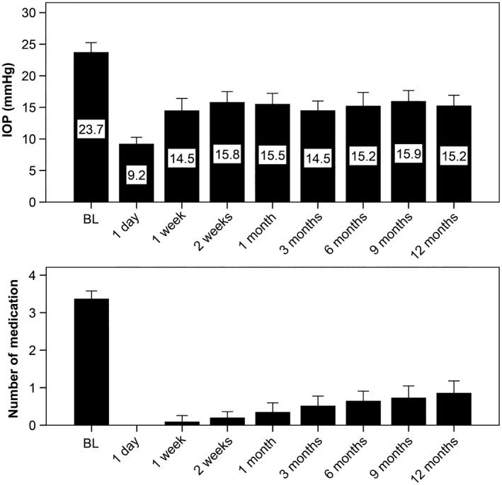
In a prospective, single-centre, single-armed cohort study, blebs were characterized using AS-OCT in 78 eyes of 60 patients at day 1, at weeks 1 and 2 and at months 1, 3, 6, 9 and 12 after XEN-GGM implantation in patients with open-angle glaucoma. Morphological bleb characteristics were correlated with IOP and surgical success.

RESULTS

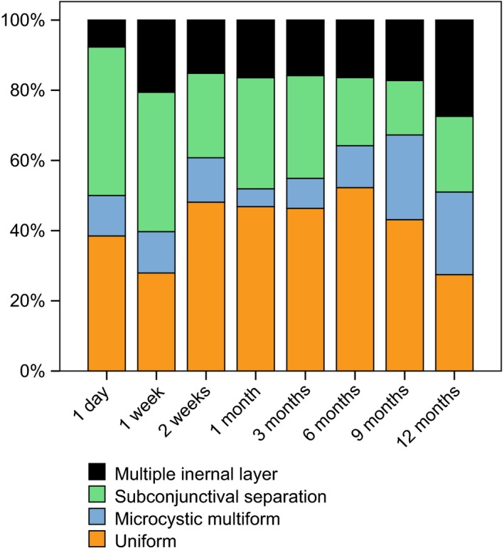
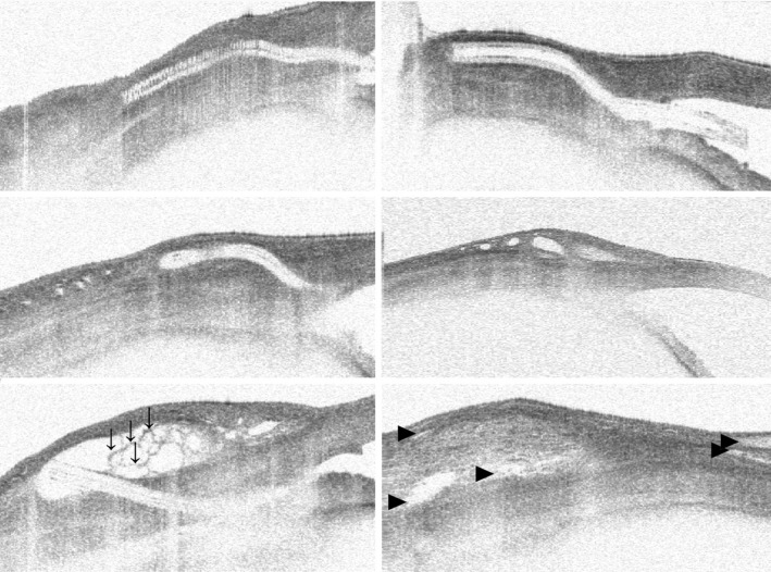
Anterior segment optical coherence tomography data indicate early and late bleb changes in the course of 12 months. Uniform blebs in AS-OCTs showed higher IOPs at all examinations between week 1 (17.7 ± 4.8 mmHg versus 11.3 ± 7.1 mmHg, p = 0.001) and month 3 (16.4 ± 6.1 versus 13.4 ± 6.1, p = 0.04). Subconjunctival tissue separation bleb morphology was associated with lower mean IOPs during the course of 12 months (r = -0.75, p = 0.031). Predictors for surgical failure at month 12 were microcystic multiform bleb morphology in AS-OCT at month 3 (60% versus 15%, relative risk 4.0, p = 0.043) and uniform bleb morphology at month 9 (33% versus 23%, relative risk 1.4, p = 0.015).

CONCLUSION

Bleb appearance after XEN surgery seems to be different to classic trabeculectomy literature. The present data suggest correlation of IOP and surgical long-term success with bleb morphology in AS-OCT. Prevalence of small diffuse cysts is directly associated with lower IOPs, while cystic encapsulation at 3 months predicts higher surgical failure.

摘要

目的

与经典小梁切除术一样,微创、内路 XEN 青光眼凝胶微支架(XEN-GGM)在结膜下形成滤过泡。本研究的目的是使用眼前节光学相干断层扫描(AS-OCT)在 XEN-GGM 植入后随时间观察内滤过泡的形态。

方法

在一项前瞻性、单中心、单臂队列研究中,在接受开角型青光眼治疗的 60 例 78 只眼中,在 XEN-GGM 植入后第 1、1、2 周、第 1、3、6、9 和 12 个月,使用 AS-OCT 对滤过泡进行特征描述。将形态滤过泡特征与 IOP 和手术成功率相关联。

结果

AS-OCT 数据表明,在 12 个月的过程中,早期和晚期滤过泡发生变化。在所有检查中,AS-OCT 中均匀的滤过泡在第 1 周(17.7±4.8mmHg 比 11.3±7.1mmHg,p=0.001)和第 3 个月(16.4±6.1mmHg 比 13.4±6.1mmHg,p=0.04)的 IOP 更高。在 12 个月的过程中,结膜下组织分离的滤过泡形态与较低的平均 IOP 相关(r=-0.75,p=0.031)。第 12 个月手术失败的预测因素为第 3 个月 AS-OCT 中小囊多形性滤过泡形态(60%比 15%,相对风险 4.0,p=0.043)和第 9 个月均匀滤过泡形态(33%比 23%,相对风险 1.4,p=0.015)。

结论

XEN 手术后的滤过泡外观似乎与经典小梁切除术文献不同。本研究数据表明,AS-OCT 中的滤过泡形态与 IOP 和手术长期成功率相关。小弥漫性囊肿的患病率与较低的 IOP 直接相关,而 3 个月时的囊性包裹则预示着较高的手术失败风险。

https://cdn.ncbi.nlm.nih.gov/pmc/blobs/2ffc/6586011/a952bfd8d475/AOS-97-e231-g004.jpg
https://cdn.ncbi.nlm.nih.gov/pmc/blobs/2ffc/6586011/32e190c7cd6d/AOS-97-e231-g001.jpg
https://cdn.ncbi.nlm.nih.gov/pmc/blobs/2ffc/6586011/d86d15d13612/AOS-97-e231-g002.jpg
https://cdn.ncbi.nlm.nih.gov/pmc/blobs/2ffc/6586011/5ff7f37998ae/AOS-97-e231-g003.jpg
https://cdn.ncbi.nlm.nih.gov/pmc/blobs/2ffc/6586011/a952bfd8d475/AOS-97-e231-g004.jpg

相似文献

1
Longitudinal bleb morphology in anterior segment OCT after minimally invasive transscleral ab interno Glaucoma Gel Microstent implantation.眼前节 OCT 下微创经巩膜内路 Glaucoma Gel Microstent 植入术后房水引流物的纵向形态。
Acta Ophthalmol. 2019 Mar;97(2):e231-e237. doi: 10.1111/aos.13902. Epub 2018 Aug 29.
2
Effect of the outer stent position on efficacy after minimally invasive transscleral glaucoma gel stent implantation.经巩膜微创青光眼凝胶支架植入术后外支架位置对疗效的影响。
Acta Ophthalmol. 2019 Dec;97(8):e1105-e1111. doi: 10.1111/aos.14167. Epub 2019 Jun 18.
3
Structural imaging of conjunctival filtering blebs in XEN gel implantation and trabeculectomy: a confocal and anterior segment optical coherence tomography study.XEN 凝胶植入术和小梁切除术的结膜滤过泡的结构成像:共焦和眼前节光学相干断层扫描研究。
Graefes Arch Clin Exp Ophthalmol. 2020 Aug;258(8):1763-1770. doi: 10.1007/s00417-020-04671-2. Epub 2020 May 15.
4
[Bleb morphology using optical coherence tomography : After primary implantation of XEN gel stent and open conjunctival revision].[使用光学相干断层扫描的泡状形态:XEN凝胶支架初次植入及开放性结膜修复术后]
Ophthalmologie. 2023 May;120(5):529-537. doi: 10.1007/s00347-022-01764-7. Epub 2022 Nov 29.
5
Morphology of functioning trabeculectomy blebs using anterior segment optical coherence tomography.使用眼前节光学相干断层扫描技术观察功能性小梁切除术滤过泡的形态学
Indian J Ophthalmol. 2014 Jun;62(6):711-4. doi: 10.4103/0301-4738.136227.
6
Efficacy of Needling Revision After XEN Gel Stent Implantation: A Prospective Study.XEN 凝胶支架植入后针刺修复的疗效:一项前瞻性研究。
J Glaucoma. 2020 Jan;29(1):11-14. doi: 10.1097/IJG.0000000000001394.
7
Imaging of trabeculectomy blebs using anterior segment optical coherence tomography.使用眼前节光学相干断层扫描对小梁切除术滤过泡进行成像。
Ophthalmology. 2007 Jan;114(1):47-53. doi: 10.1016/j.ophtha.2006.05.078. Epub 2006 Oct 27.
8
Four-year results of a minimally invasive transscleral glaucoma gel stent implantation in a prospective multi-centre study.一项前瞻性多中心研究中经巩膜微创青光眼凝胶支架植入的 4 年结果。
Clin Exp Ophthalmol. 2019 Jul;47(5):581-587. doi: 10.1111/ceo.13463. Epub 2019 Feb 3.
9
Two-year outcomes of minimally invasive XEN Gel Stent implantation in primary open-angle and pseudoexfoliation glaucoma.原发性开角型青光眼和假性剥脱性青光眼微创 XEN 凝胶支架植入的两年疗效。
Acta Ophthalmol. 2021 Jun;99(4):369-375. doi: 10.1111/aos.14627. Epub 2020 Sep 30.
10
Optical coherence tomography analysis of filtering blebs after long-term, functioning trabeculectomy and XEN® stent implant.长期功能性小梁切除术及XEN®支架植入术后滤过泡的光学相干断层扫描分析
Graefes Arch Clin Exp Ophthalmol. 2019 May;257(5):1005-1011. doi: 10.1007/s00417-019-04272-8. Epub 2019 Feb 20.

引用本文的文献

1
Surgical Outcomes of XEN45 Gel Stent Using Ab Interno Technique in Open-Angle Glaucoma: A 2-Year Follow-Up Study.采用内路技术植入XEN45凝胶支架治疗开角型青光眼的手术效果:一项为期2年的随访研究。
J Clin Med. 2025 Jun 30;14(13):4617. doi: 10.3390/jcm14134617.
2
Discriminatory power of trabeculectomy bleb internal reflectivity and morphology in surgical success using anterior segment optical coherence tomography.使用眼前节光学相干断层扫描技术评估小梁切除术滤过泡内部反射率和形态对手术成功的鉴别能力。
BMC Ophthalmol. 2025 Jan 30;25(1):52. doi: 10.1186/s12886-024-03770-6.
3
Visualization of Scleral Flap Patency in Glaucoma Filtering Blebs Using OCT.

本文引用的文献

1
Efficacy, Safety, and Risk Factors for Failure of Standalone Ab Interno Gelatin Microstent Implantation versus Standalone Trabeculectomy.单纯经内路巩膜凝胶微支架植入术与单纯小梁切除术的疗效、安全性和失败的风险因素。
Ophthalmology. 2017 Nov;124(11):1579-1588. doi: 10.1016/j.ophtha.2017.05.004. Epub 2017 Jun 7.
2
Glaucoma treatment trends: a review.青光眼治疗趋势:综述
Can J Ophthalmol. 2017 Feb;52(1):114-124. doi: 10.1016/j.jcjo.2016.07.013. Epub 2016 Nov 17.
3
Fluid Dynamics of a Novel Micro-Fistula Implant for the Surgical Treatment of Glaucoma.
使用光学相干断层扫描(OCT)观察青光眼滤过泡中巩膜瓣的通畅情况。
Ophthalmol Sci. 2024 Aug 23;5(1):100604. doi: 10.1016/j.xops.2024.100604. eCollection 2025 Jan-Feb.
4
Superonasal vs Inferonasal Subconjunctival Gel Stent Placement in Patients with Glaucoma.青光眼患者超鼻侧与鼻下侧结膜下凝胶支架植入的比较
J Curr Glaucoma Pract. 2024 Apr-Jun;18(2):63-67. doi: 10.5005/jp-journals-10078-1441. Epub 2024 Jul 10.
5
Anterior-Segment Optical Coherence Tomography-Based Evaluation of the Journey of a Bleb in the Early Postoperative Period.基于眼前节光学相干断层扫描的术后早期滤过泡行程评估
Cureus. 2024 Jun 30;16(6):e63512. doi: 10.7759/cureus.63512. eCollection 2024 Jun.
6
XEN45 Implant in Medically Controlled vs. Uncontrolled Eyes-Differential IOP Changes in Real-Life Conditions.XEN45植入物在医学控制与未控制的眼睛中的应用——现实生活条件下的眼压差异变化
J Clin Med. 2024 Jun 11;13(12):3406. doi: 10.3390/jcm13123406.
7
VEGF-C and 5-Fluorouracil Improve Bleb Survival in a Rabbit Glaucoma Surgery Trabeculectomy Model.VEGF-C 和 5-氟尿嘧啶可提高兔青光眼手术小梁切除术模型中的滤过泡存活率。
Invest Ophthalmol Vis Sci. 2024 May 1;65(5):32. doi: 10.1167/iovs.65.5.32.
8
Bleb Morphology on Anterior-Segment Optical Coherence Tomography after XEN Gel Stent Implantation.XEN凝胶支架植入术后眼前节光学相干断层扫描的泡状形态
J Clin Med. 2023 Oct 25;12(21):6740. doi: 10.3390/jcm12216740.
9
Comparison of Bleb Morphology following PRESERFLO MicroShunt and Trabeculectomy Using Anterior Segment OCT.使用眼前节光学相干断层扫描(OCT)比较 PRESERFLO 微分流术和小梁切除术后的滤过泡形态
Diagnostics (Basel). 2023 Nov 2;13(21):3373. doi: 10.3390/diagnostics13213373.
10
Swept-Source Anterior Segment Optical Coherence Tomography Imaging and Quantification of Bleb Parameters in Glaucoma Filtration Surgery.扫频源眼前节光学相干断层扫描成像及青光眼滤过手术中滤过泡参数的量化分析
Bioengineering (Basel). 2023 Oct 13;10(10):1186. doi: 10.3390/bioengineering10101186.
用于青光眼手术治疗的新型微瘘植入物的流体动力学
Invest Ophthalmol Vis Sci. 2015 Jul;56(8):4789-95. doi: 10.1167/iovs.15-16625.
4
Lymphatic and Blood Vessel Density in Human Conjunctiva After Glaucoma Filtration Surgery.青光眼滤过术后人结膜的淋巴管和血管密度
J Glaucoma. 2016 Jan;25(1):e35-8. doi: 10.1097/IJG.0000000000000199.
5
Anterior segment optical coherence tomography imaging of conjunctival filtering blebs after glaucoma surgery.青光眼手术后结膜滤过泡的眼前节光学相干断层扫描成像
Biomed Res Int. 2014;2014:610623. doi: 10.1155/2014/610623. Epub 2014 Jul 20.
6
[Long-term results after trabeculectomy with 5-fluorouracil].[5-氟尿嘧啶小梁切除术后的长期结果]
Ophthalmologe. 2014 Aug;111(8):749-56. doi: 10.1007/s00347-013-2970-3.
7
Mitomycin C versus 5-fluorouracil as an adjunctive treatment for trabeculectomy: a meta-analysis of randomized clinical trials.丝裂霉素 C 与 5-氟尿嘧啶作为小梁切除术的辅助治疗:随机临床试验的荟萃分析。
Clin Exp Ophthalmol. 2013 Nov;41(8):798-806. doi: 10.1111/ceo.12097.
8
Trabeculectomy in the 21st century: a multicenter analysis.21 世纪的小梁切除术:一项多中心分析。
Ophthalmology. 2013 Dec;120(12):2532-2539. doi: 10.1016/j.ophtha.2013.07.049. Epub 2013 Sep 23.
9
Optical coherence tomography of the suprachoroid after CyPass Micro-Stent implantation for the treatment of open-angle glaucoma.脉络膜上腔光相干断层扫描在 CyPass Micro-Stent 植入术后治疗开角型青光眼的应用。
Br J Ophthalmol. 2014 Jan;98(1):19-23. doi: 10.1136/bjophthalmol-2012-302951. Epub 2013 Jun 6.
10
Micro-invasive glaucoma surgery: current perspectives and future directions.微创青光眼手术:现状与未来方向。
Curr Opin Ophthalmol. 2012 Mar;23(2):96-104. doi: 10.1097/ICU.0b013e32834ff1e7.