Choudhary Tanvi S, Raval Reema M, Shah Kintu S, Gajwani Sakshi M, Mehta Radha J, Patel Megha C
Glaucoma and Squint Clinic, Shri Chimanlal Harilal (CH) Nagri Municipal Eye Hospital, Ahmedabad, IND.
Department of Psychiatry, Gujarat Medical Education and Research Society (GMERS) Medical College and Hospital, Navsari, IND.
Cureus. 2024 Jun 30;16(6):e63512. doi: 10.7759/cureus.63512. eCollection 2024 Jun.
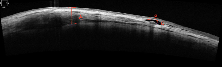
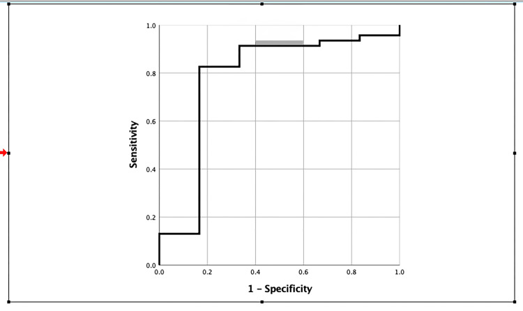
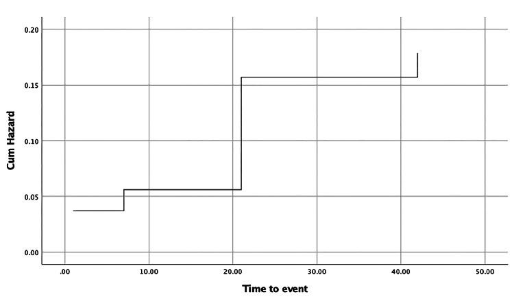
Introduction In the early postoperative period following trabeculectomy, monitoring the journey of bleb formation is crucial for assessing surgical success. Anterior-segment optical coherence tomography (AS-OCT) emerges as a powerful tool in this pursuit, offering high-resolution imaging of bleb morphology and dynamics. This study aims to evaluate the internal structure of blebs through their maturation phases using AS-OCT. Methods Fifty-five eyes undergoing trabeculectomy were enrolled in a prospective observational study. Serial AS-OCT examinations were done on day 1, week 1, week 3, and week 6 postoperatively; bleb parameters were calculated and correlated with intraocular pressure (IOP). Results IOP control was seen in 45 eyes six months of post-trabeculectomy. Multiform bleb wall reflectivity (BWR) statistically correlates with the success of trabeculectomy. Blebs were successful if BWR showed no change from day 1 to week 6. BWR remained the same on all follow-ups if week 1 bleb wall thickness (BWT) was less than 129.5 microns with 82.6% sensitivity and 83.3% specificity. The cumulative hazard of change in BWR is estimated to be approximately 5.6%, 15.7%, and 17.9% at week 1, week 3, and week 6 follow-ups, respectively. Conclusions Successful blebs showed consistent BWR from day 1 to week 6 of follow-up. Serial AS-OCT examination for changes in BWR in early stages can be done to predict the fate of bleb. The maximum change in BWR occurs between the week 1 and week 3 follow-up periods requiring close follow-up.
引言 在小梁切除术后的早期,监测滤过泡形成过程对于评估手术成功率至关重要。眼前节光学相干断层扫描(AS-OCT)成为实现这一目标的有力工具,可提供滤过泡形态和动态的高分辨率成像。本研究旨在使用AS-OCT评估滤过泡在其成熟阶段的内部结构。
方法 55只接受小梁切除术的眼睛纳入一项前瞻性观察性研究。在术后第1天、第1周、第3周和第6周进行系列AS-OCT检查;计算滤过泡参数并与眼压(IOP)相关联。
结果 小梁切除术后6个月,45只眼睛眼压得到控制。多形性滤过泡壁反射率(BWR)与小梁切除术的成功率具有统计学相关性。如果BWR从第1天到第6周无变化,则滤过泡手术成功。如果第1周滤过泡壁厚度(BWT)小于129.5微米,所有随访中BWR保持不变,敏感性为82.6%,特异性为83.3%。在第1周、第3周和第6周随访时,BWR变化的累积风险估计分别约为5.6%、15.7%和17.9%。
结论 成功的滤过泡在随访的第1天到第6周显示出一致的BWR。可进行系列AS-OCT检查早期BWR的变化以预测滤过泡的转归。BWR的最大变化发生在第1周和第3周随访期间,需要密切随访。