• 文献检索
  • 文档翻译
  • 深度研究
  • 学术资讯
  • Suppr Zotero 插件Zotero 插件
  • 邀请有礼
  • 套餐&价格
  • 历史记录
应用&插件
Suppr Zotero 插件Zotero 插件浏览器插件Mac 客户端Windows 客户端微信小程序
定价
高级版会员购买积分包购买API积分包
服务
文献检索文档翻译深度研究API 文档MCP 服务
关于我们
关于 Suppr公司介绍联系我们用户协议隐私条款
关注我们

Suppr 超能文献

核心技术专利:CN118964589B侵权必究
粤ICP备2023148730 号-1Suppr @ 2026

文献检索

告别复杂PubMed语法,用中文像聊天一样搜索,搜遍4000万医学文献。AI智能推荐,让科研检索更轻松。

立即免费搜索

文件翻译

保留排版,准确专业,支持PDF/Word/PPT等文件格式,支持 12+语言互译。

免费翻译文档

深度研究

AI帮你快速写综述,25分钟生成高质量综述,智能提取关键信息,辅助科研写作。

立即免费体验

角膜形态发生过程中胶原纤维宏观结构的细胞调控。

Cell regulation of collagen fibril macrostructure during corneal morphogenesis.

机构信息

Gavin Herbert Eye Institute, University of California, Irvine, Irvine, CA, USA; Structural Biophysics Research Group, School of Optometry and Vision Sciences, Cardiff University, Cardiff, Wales, UK.

Gavin Herbert Eye Institute, University of California, Irvine, Irvine, CA, USA.

出版信息

Acta Biomater. 2018 Oct 1;79:96-112. doi: 10.1016/j.actbio.2018.08.017. Epub 2018 Aug 29.

DOI:10.1016/j.actbio.2018.08.017
PMID:30170195
原文链接:https://pmc.ncbi.nlm.nih.gov/articles/PMC6163070/
Abstract

UNLABELLED

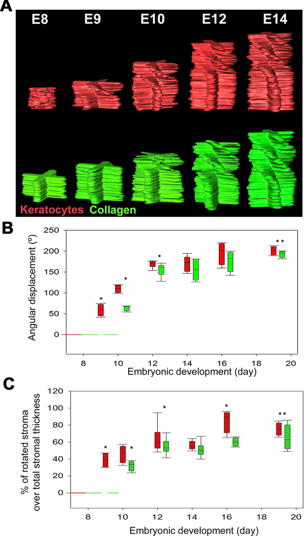
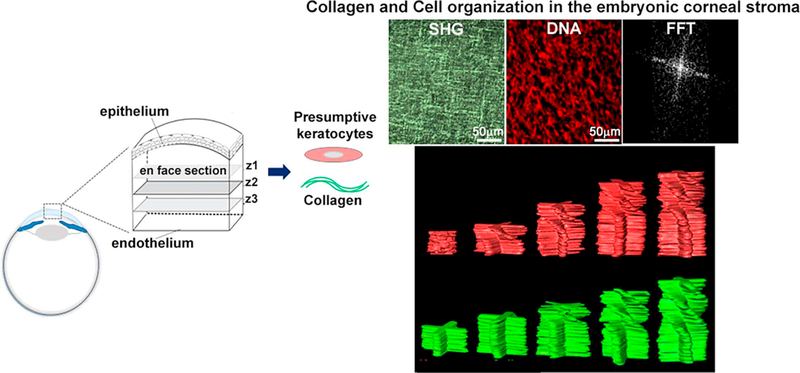
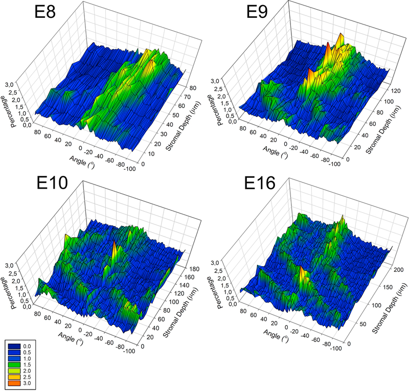
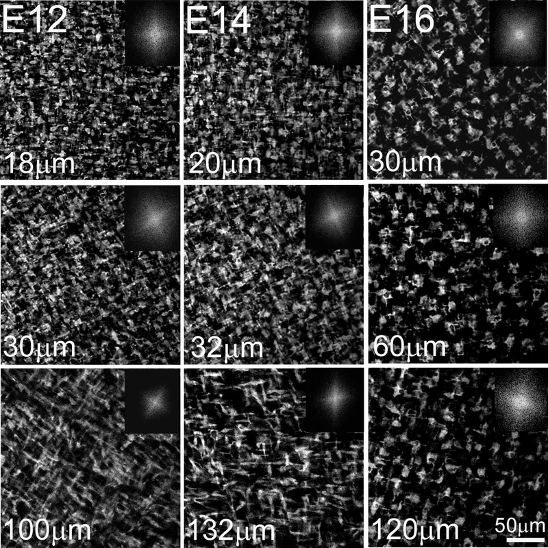
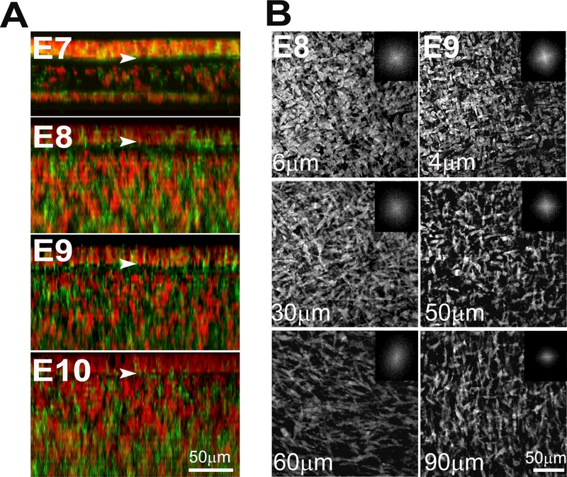
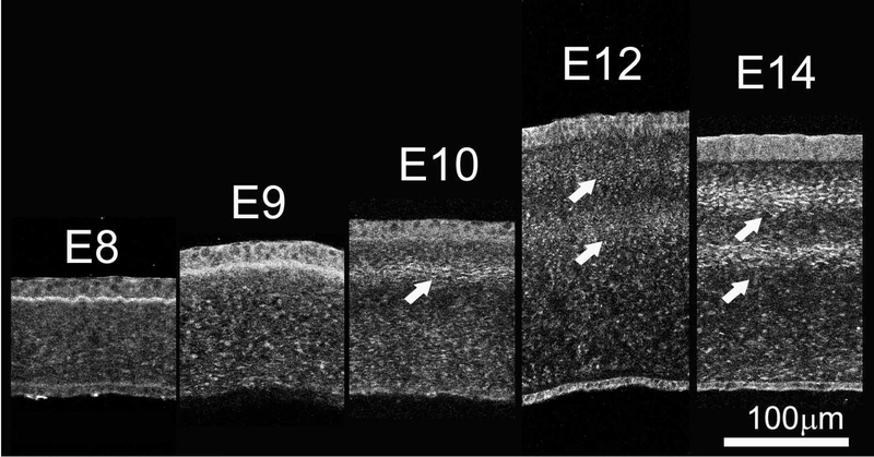
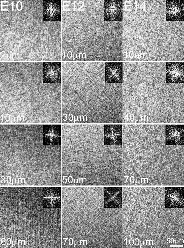
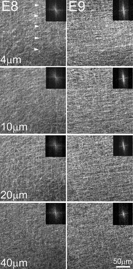
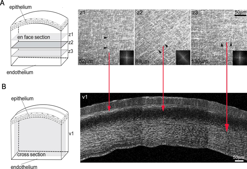
While tissue form and function is highly dependent upon tissue-specific collagen composition and organization, little is known of the mechanisms controlling the bundling of collagen fibrils into fibers and larger structural designs that lead to the formation of bones, tendons and other tissues. Using the cornea as a model system, our previous 3 dimensional mapping of collagen fiber organization has demonstrated that macrostructural organization of collagen fibers involving interweaving, branching and anastomosing plays a critical role in controlling mechanical stiffness, corneal shape and refractive power. In this work, the cellular and mechanical mechanisms regulating critical events in the assembly of collagen macrostructure are analysed in the developing chicken cornea. We elucidated the temporal events leading to adult corneal structure and determined the effects of intraocular pressure (IOP) on the organization of the collagen macrostructure. Our findings indicate that the complex adult collagen organization begins to appear on embryonic day 10 (E10) after deposition of the primary stroma and full invasion of keratocytes. Importantly, organizational changes in keratocytes appearing at E9 preceded and predicted later changes in collagen organization. Corneal collagen organization remained unaffected when the development of IOP was blocked at E4. These findings support a primary role for keratocytes in controlling stromal organization, mechanical stiffness and corneal shape that are not regulated by the IOP. Our findings also suggest that the avian cornea represents an excellent experimental model for elucidating key regulatory steps and mechanisms controlling the collagen fiber organization that is critical to determining tissue form and function.

STATEMENT OF SIGNIFICANCE

This work by using an ex ovo model system, begins to investigate the potential mechanisms controlling collagen fibril macrostructure. In particular, this work highlights a convergent role for the corneal keratocytes in organizing the complex collagen macrostructure, necessary to support high visual acuity. Our data supports that the intraocular pressure does not influence collagen fibril macrostructure and suggest that the avian cornea represents an excellent experimental model for elucidating key regulatory steps and mechanisms controlling the collagen fiber organization that is critical to determining tissue form and function. Clearly understanding the cellular and molecular mechanisms that underlie collagen fibril macrostructure will be highly beneficial for future tissue engineering and regenerative medicine applications.

摘要

未加标签

虽然组织的形态和功能高度依赖于组织特异性胶原蛋白的组成和组织,但对于控制胶原蛋白原纤维束成纤维和更大结构设计的机制知之甚少,这些结构设计导致骨骼、肌腱和其他组织的形成。我们以前使用角膜作为模型系统,对胶原纤维组织的三维图谱进行了研究,结果表明,涉及交织、分支和吻合的胶原纤维的宏观结构组织对于控制机械刚度、角膜形状和屈光力起着关键作用。在这项工作中,分析了发育中的鸡角膜中调节胶原大分子组装的关键事件的细胞和机械机制。我们阐明了导致成年角膜结构的时间事件,并确定了眼内压(IOP)对胶原大分子结构的影响。我们的研究结果表明,复杂的成年胶原组织在胚胎第 10 天(E10 天)初级基质沉积和角膜细胞完全浸润后开始出现。重要的是,E9 天出现的角膜细胞的组织变化先于并预测了胶原组织的后期变化。当 E4 天阻断 IOP 发育时,角膜胶原组织不受影响。这些发现支持角膜细胞在控制基质组织、机械刚度和角膜形状方面发挥主要作用,而这些不受 IOP 调节。我们的研究结果还表明,禽类角膜是阐明控制对确定组织形态和功能至关重要的胶原纤维组织的关键调节步骤和机制的优秀实验模型。

https://cdn.ncbi.nlm.nih.gov/pmc/blobs/be40/6163070/674183dd18a7/nihms-1505522-f0011.jpg
https://cdn.ncbi.nlm.nih.gov/pmc/blobs/be40/6163070/1bdd5a9ad3db/nihms-1505522-f0001.jpg
https://cdn.ncbi.nlm.nih.gov/pmc/blobs/be40/6163070/0f5963fb7c13/nihms-1505522-f0002.jpg
https://cdn.ncbi.nlm.nih.gov/pmc/blobs/be40/6163070/079af7c2b087/nihms-1505522-f0003.jpg
https://cdn.ncbi.nlm.nih.gov/pmc/blobs/be40/6163070/1f739f43a4ae/nihms-1505522-f0004.jpg
https://cdn.ncbi.nlm.nih.gov/pmc/blobs/be40/6163070/b326d92911b7/nihms-1505522-f0005.jpg
https://cdn.ncbi.nlm.nih.gov/pmc/blobs/be40/6163070/acad1a0c0ebf/nihms-1505522-f0006.jpg
https://cdn.ncbi.nlm.nih.gov/pmc/blobs/be40/6163070/a9ff970ba03b/nihms-1505522-f0007.jpg
https://cdn.ncbi.nlm.nih.gov/pmc/blobs/be40/6163070/a42122ae252c/nihms-1505522-f0008.jpg
https://cdn.ncbi.nlm.nih.gov/pmc/blobs/be40/6163070/6e704d6f7dc4/nihms-1505522-f0009.jpg
https://cdn.ncbi.nlm.nih.gov/pmc/blobs/be40/6163070/e625a77b26da/nihms-1505522-f0010.jpg
https://cdn.ncbi.nlm.nih.gov/pmc/blobs/be40/6163070/674183dd18a7/nihms-1505522-f0011.jpg

相似文献

1
Cell regulation of collagen fibril macrostructure during corneal morphogenesis.角膜形态发生过程中胶原纤维宏观结构的细胞调控。
Acta Biomater. 2018 Oct 1;79:96-112. doi: 10.1016/j.actbio.2018.08.017. Epub 2018 Aug 29.
2
Shear-induced alignment of collagen fibrils using 3D cell printing for corneal stroma tissue engineering.使用 3D 细胞打印对胶原蛋白纤维进行切变诱导排列以用于角膜基质组织工程。
Biofabrication. 2019 May 7;11(3):035017. doi: 10.1088/1758-5090/ab1a8b.
3
Cell-independent matrix configuration in early corneal development.早期角膜发育中的细胞非依赖性基质构象。
Exp Eye Res. 2019 Oct;187:107772. doi: 10.1016/j.exer.2019.107772. Epub 2019 Aug 21.
4
Collagen fibril assembly in the developing avian primary corneal stroma.发育中的鸡胚初级角膜基质中的胶原原纤维组装
Invest Ophthalmol Vis Sci. 1994 Mar;35(3):862-9.
5
Spatial and temporal alterations in the collagen fibrillar array during the onset of transparency in the avian cornea.鸟类角膜透明化开始过程中胶原纤维阵列的时空变化。
Exp Eye Res. 2004 May;78(5):909-15. doi: 10.1016/j.exer.2004.01.005.
6
Regulation of corneal stroma extracellular matrix assembly.角膜基质细胞外基质组装的调控
Exp Eye Res. 2015 Apr;133:69-80. doi: 10.1016/j.exer.2014.08.001.
7
Keratan sulfate glycosaminoglycan and the association with collagen fibrils in rudimentary lamellae in the developing avian cornea.硫酸角质素糖胺聚糖及其与发育中的鸡角膜原始板层中胶原纤维的关联。
Invest Ophthalmol Vis Sci. 2007 Jul;48(7):3083-8. doi: 10.1167/iovs.06-1323.
8
The morphogenesis of the chick primary corneal stroma. I. New observations on collagen organization in vivo help explain stromal deposition and growth.鸡原发性角膜基质的形态发生。I. 关于体内胶原蛋白组织的新观察有助于解释基质沉积和生长。
Development. 1987 May;100(1):135-45. doi: 10.1242/dev.100.1.135.
9
Formation of stromal collagen fibrils and proteoglycans in the developing zebrafish cornea.斑马鱼发育中角膜基质胶原纤维和蛋白聚糖的形成。
Acta Ophthalmol. 2008 Sep;86(6):655-65. doi: 10.1111/j.1600-0420.2007.01135.x.
10
Human corneal fibrillogenesis. Collagen V structural analysis and fibrillar assembly by stromal fibroblasts in culture.人角膜纤维生成。培养的基质成纤维细胞对V型胶原蛋白的结构分析及纤维组装
Invest Ophthalmol Vis Sci. 1996 Aug;37(9):1749-60.

引用本文的文献

1
A bioequivalent cornea cross-linking method using photo-initiators LAP and visible light.一种使用光引发剂LAP和可见光的生物等效性角膜交联方法。
Mater Today Bio. 2025 Jul 17;34:102110. doi: 10.1016/j.mtbio.2025.102110. eCollection 2025 Oct.
2
Dimensionality Matters: Exploiting UV-Photopatterned 2D and Two-Photon-Printed 2.5D Contact Guidance Cues to Control Corneal Fibroblast Behavior and Collagen Deposition.维度很重要:利用紫外光图案化的二维和双光子打印的2.5维接触引导线索来控制角膜成纤维细胞行为和胶原蛋白沉积。
Bioengineering (Basel). 2024 Apr 19;11(4):402. doi: 10.3390/bioengineering11040402.
3
Developmental Changes in Patterns of Distribution of Fibronectin and Tenascin-C in the Chicken Cornea: Evidence for Distinct and Independent Functions during Corneal Development and Morphogenesis.

本文引用的文献

1
Evolution of the vertebrate corneal stroma.脊椎动物角膜基质的演化。
Prog Retin Eye Res. 2018 May;64:65-76. doi: 10.1016/j.preteyeres.2018.01.002. Epub 2018 Feb 2.
2
Tissue and cellular biomechanics during corneal wound injury and repair.角膜伤口损伤与修复过程中的组织和细胞生物力学
Acta Biomater. 2017 Aug;58:291-301. doi: 10.1016/j.actbio.2017.05.051. Epub 2017 May 27.
3
Human Corneal Fibroblast Pattern Evolution and Matrix Synthesis on Mechanically Biased Substrates.人角膜成纤维细胞在机械偏置基质上的形态演变及基质合成
鸡角膜中纤维连接蛋白和腱糖蛋白-C分布模式的发育变化:角膜发育和形态发生过程中具有独特和独立功能的证据。
Int J Mol Sci. 2023 Feb 10;24(4):3555. doi: 10.3390/ijms24043555.
4
Chondroitin Sulphate/Dermatan Sulphate Proteoglycans: Potential Regulators of Corneal Stem/Progenitor Cell Phenotype In Vitro.硫酸软骨素/硫酸皮肤素蛋白聚糖:体外角膜干/祖细胞表型的潜在调控因子。
Int J Mol Sci. 2023 Jan 20;24(3):2095. doi: 10.3390/ijms24032095.
5
Recapitulation of normal collagen architecture in embryonic wounded corneas.胚胎期受创角膜中正常胶原结构的概述。
Sci Rep. 2020 Aug 14;10(1):13815. doi: 10.1038/s41598-020-70658-y.
6
Identification of a Primary Stroma and Novel Endothelial Cell Projections in the Developing Human Cornea.鉴定人眼角膜发育过程中的初级基质和新型内皮细胞突起。
Invest Ophthalmol Vis Sci. 2020 Jun 3;61(6):5. doi: 10.1167/iovs.61.6.5.
7
Cell-independent matrix configuration in early corneal development.早期角膜发育中的细胞非依赖性基质构象。
Exp Eye Res. 2019 Oct;187:107772. doi: 10.1016/j.exer.2019.107772. Epub 2019 Aug 21.
8
Three-dimensional imaging of the extracellular matrix and cell interactions in the developing prenatal mouse cornea.发育中胚胎期小鼠角膜细胞外基质和细胞相互作用的三维成像。
Sci Rep. 2019 Aug 2;9(1):11277. doi: 10.1038/s41598-019-47653-z.
9
Quantification of collagen fiber structure using second harmonic generation imaging and two-dimensional discrete Fourier transform analysis: Application to the human optic nerve head.利用二次谐波产生成像和二维离散傅里叶变换分析对胶原纤维结构进行定量分析:在人视神经乳头中的应用。
J Biophotonics. 2019 May;12(5):e201800376. doi: 10.1002/jbio.201800376. Epub 2019 Jan 10.
Tissue Eng Part A. 2016 Oct;22(19-20):1204-1217. doi: 10.1089/ten.TEA.2016.0164. Epub 2016 Sep 29.
4
Corneal Fibroblast Migration Patterns During Intrastromal Wound Healing Correlate With ECM Structure and Alignment.基质内伤口愈合过程中角膜成纤维细胞迁移模式与细胞外基质结构及排列相关。
Invest Ophthalmol Vis Sci. 2015 Nov;56(12):7352-61. doi: 10.1167/iovs.15-17978.
5
Corneal structure and transparency.角膜结构与透明度。
Prog Retin Eye Res. 2015 Nov;49:1-16. doi: 10.1016/j.preteyeres.2015.07.001. Epub 2015 Jul 2.
6
A Comparative Study of Vertebrate Corneal Structure: The Evolution of a Refractive Lens.脊椎动物角膜结构的比较研究:屈光晶状体的进化
Invest Ophthalmol Vis Sci. 2015 Apr;56(4):2764-72. doi: 10.1167/iovs.15-16584.
7
Regulation of corneal stroma extracellular matrix assembly.角膜基质细胞外基质组装的调控
Exp Eye Res. 2015 Apr;133:69-80. doi: 10.1016/j.exer.2014.08.001.
8
Optimized ex-ovo culturing of chick embryos to advanced stages of development.将鸡胚的体外培养优化至发育晚期阶段。
J Vis Exp. 2015 Jan 24(95):52129. doi: 10.3791/52129.
9
A role for topographic cues in the organization of collagenous matrix by corneal fibroblasts and stem cells.在角膜成纤维细胞和干细胞胶原基质的组织中,地形线索的作用。
PLoS One. 2014 Jan 21;9(1):e86260. doi: 10.1371/journal.pone.0086260. eCollection 2014.
10
Three-dimensional aspects of matrix assembly by cells in the developing cornea.细胞在发育中的角膜中进行基质组装的三维方面。
Proc Natl Acad Sci U S A. 2014 Jan 14;111(2):687-92. doi: 10.1073/pnas.1313561110. Epub 2014 Jan 2.