• 文献检索
  • 文档翻译
  • 深度研究
  • 学术资讯
  • Suppr Zotero 插件Zotero 插件
  • 邀请有礼
  • 套餐&价格
  • 历史记录
应用&插件
Suppr Zotero 插件Zotero 插件浏览器插件Mac 客户端Windows 客户端微信小程序
定价
高级版会员购买积分包购买API积分包
服务
文献检索文档翻译深度研究API 文档MCP 服务
关于我们
关于 Suppr公司介绍联系我们用户协议隐私条款
关注我们

Suppr 超能文献

核心技术专利:CN118964589B侵权必究
粤ICP备2023148730 号-1Suppr @ 2026

文献检索

告别复杂PubMed语法,用中文像聊天一样搜索,搜遍4000万医学文献。AI智能推荐,让科研检索更轻松。

立即免费搜索

文件翻译

保留排版,准确专业,支持PDF/Word/PPT等文件格式,支持 12+语言互译。

免费翻译文档

深度研究

AI帮你快速写综述,25分钟生成高质量综述,智能提取关键信息,辅助科研写作。

立即免费体验

前列腺癌的计算扩散加权成像:基础、优势、注意事项和未来展望。

Computed Diffusion-Weighted Imaging in Prostate Cancer: Basics, Advantages, Cautions, and Future Prospects.

机构信息

Department of Radiology, Kobe University Graduate School of Medicine, Kobe 650-0017, Japan.

Department of Radiology, Kawasaki Medical School, Kurashiki 701-0192, Japan.

出版信息

Korean J Radiol. 2018 Sep-Oct;19(5):832-837. doi: 10.3348/kjr.2018.19.5.832. Epub 2018 Aug 6.

DOI:10.3348/kjr.2018.19.5.832
PMID:30174471
原文链接:https://pmc.ncbi.nlm.nih.gov/articles/PMC6082756/
Abstract

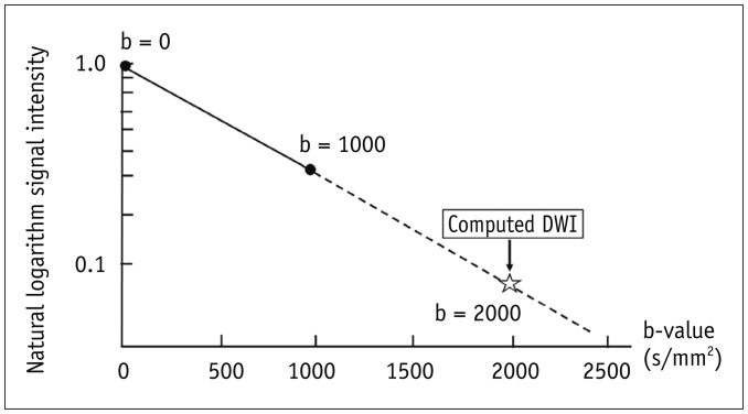
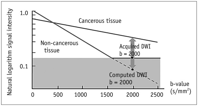
Computed diffusion-weighted MRI is a recently proposed post-processing technique that produces b-value images from diffusion-weighted imaging (DWI), acquired using at least two different b-values. This article presents an argument for computed DWI for prostate cancer by viewing four aspects of DWI: fundamentals, image quality and diagnostic performance, computing procedures, and future uses.

摘要

计算扩散加权磁共振成像(DWI)是一种最近提出的后处理技术,它可以从使用至少两个不同 b 值采集的扩散加权成像(DWI)中生成 b 值图像。本文从 DWI 的四个方面(基本原理、图像质量和诊断性能、计算过程以及未来用途)来看待前列腺癌的计算 DWI,提出了一个论点。

https://cdn.ncbi.nlm.nih.gov/pmc/blobs/f926/6082756/07a733317b29/kjr-19-832-g004.jpg
https://cdn.ncbi.nlm.nih.gov/pmc/blobs/f926/6082756/26c2e0265d8d/kjr-19-832-g001.jpg
https://cdn.ncbi.nlm.nih.gov/pmc/blobs/f926/6082756/431bcd5ee246/kjr-19-832-g002.jpg
https://cdn.ncbi.nlm.nih.gov/pmc/blobs/f926/6082756/cfffac86618b/kjr-19-832-g003.jpg
https://cdn.ncbi.nlm.nih.gov/pmc/blobs/f926/6082756/07a733317b29/kjr-19-832-g004.jpg

相似文献

1
Computed Diffusion-Weighted Imaging in Prostate Cancer: Basics, Advantages, Cautions, and Future Prospects.前列腺癌的计算扩散加权成像:基础、优势、注意事项和未来展望。
Korean J Radiol. 2018 Sep-Oct;19(5):832-837. doi: 10.3348/kjr.2018.19.5.832. Epub 2018 Aug 6.
2
Computed diffusion-weighted imaging of the prostate at 3 T: impact on image quality and tumour detection.3T 磁共振成像下前列腺的扩散加权成像:对图像质量和肿瘤检测的影响
Eur Radiol. 2013 Nov;23(11):3170-7. doi: 10.1007/s00330-013-2917-8. Epub 2013 Jun 12.
3
High and ultra-high b-value diffusion-weighted imaging in prostate cancer: a quantitative analysis.前列腺癌的高b值和超高b值扩散加权成像:定量分析
Acta Radiol. 2015 Aug;56(8):1009-15. doi: 10.1177/0284185114547900. Epub 2014 Aug 28.
4
Evaluation of Exponential ADC (eADC) and Computed DWI (cDWI) for the Detection of Prostate Cancer.评估指数型扩散加权成像(eADC)和计算扩散加权成像(cDWI)在前列腺癌检测中的应用。
Rofo. 2018 Aug;190(8):758-766. doi: 10.1055/a-0637-9980. Epub 2018 Jul 25.
5
Diffusion-weighted imaging in the assessment of prostate cancer: Comparison of zoomed imaging and conventional technique.扩散加权成像在前列腺癌评估中的应用:放大成像与传统技术的比较。
Eur J Radiol. 2016 May;85(5):893-900. doi: 10.1016/j.ejrad.2016.02.020. Epub 2016 Feb 27.
6
Accuracy of high b-value diffusion-weighted MRI for prostate cancer detection: a meta-analysis.高b值扩散加权磁共振成像用于前列腺癌检测的准确性:一项荟萃分析。
Acta Radiol. 2018 Jan;59(1):105-113. doi: 10.1177/0284185117702181. Epub 2017 Apr 4.
7
Anterior Prostate Cancer: Diagnostic Performance of T2-Weighted MRI and an Apparent Diffusion Coefficient Map.前列腺癌的前部:T2 加权 MRI 和表观扩散系数图的诊断性能。
AJR Am J Roentgenol. 2015 Aug;205(2):W185-92. doi: 10.2214/AJR.14.13392.
8
The impact of computed high b-value images on the diagnostic accuracy of DWI for prostate cancer: A receiver operating characteristics analysis.计算高 b 值图像对前列腺癌 DWI 诊断准确性的影响:一项受试者工作特征分析。
Sci Rep. 2018 Feb 21;8(1):3409. doi: 10.1038/s41598-018-21523-6.
9
Quantitative evaluation of computed high B value diffusion-weighted magnetic resonance imaging of the prostate.前列腺计算机高B值扩散加权磁共振成像的定量评估
Invest Radiol. 2013 Nov;48(11):779-86. doi: 10.1097/RLI.0b013e31829705bb.
10
Detecting prostate cancer and prostatic calcifications using advanced magnetic resonance imaging.使用先进的磁共振成像检测前列腺癌和前列腺钙化。
Asian J Androl. 2017 Jul-Aug;19(4):439-443. doi: 10.4103/1008-682X.177840.

引用本文的文献

1
Toward a Refined PI-RADS: The Feasibility and Limitations of More Informative Metrics in Reviewing MRI Scans.迈向优化的前列腺影像报告和数据系统(PI-RADS):在MRI扫描评估中采用更多信息性指标的可行性与局限性
J Magn Reson Imaging. 2025 Sep;62(3):673-690. doi: 10.1002/jmri.29754. Epub 2025 Mar 26.
2
High-resolution Diffusion-weighted Imaging of the Prostate Using Multiplexed Sensitivity-encoding: Comparison with the Conventional and Reduced Field-of-view Techniques.使用多重灵敏度编码的前列腺高分辨率扩散加权成像:与传统和缩小视野技术的比较。
Magn Reson Med Sci. 2025 Jan 1;24(1):58-65. doi: 10.2463/mrms.mp.2023-0039. Epub 2023 Oct 28.
3

本文引用的文献

1
Reduced field-of-view diffusion-weighted MRI in patients with cervical cancer.宫颈癌患者的缩小视野扩散加权磁共振成像
Br J Radiol. 2018 Jul;91(1087):20170864. doi: 10.1259/bjr.20170864. Epub 2018 Apr 16.
2
PI-RADS version 2: evaluation of diffusion-weighted imaging interpretation between b = 1000 and b = 1500 s mm.前列腺影像报告和数据系统(PI-RADS)第2版:b值为1000和1500 s/mm²时扩散加权成像解读的评估
Br J Radiol. 2017 Nov;90(1079):20170438. doi: 10.1259/bjr.20170438. Epub 2017 Oct 6.
3
Utility of computed diffusion-weighted MRI for predicting aggressiveness of prostate cancer.
Acquisition Parameters Influence Diffusion Metrics Effectiveness in Probing Prostate Tumor and Age-Related Microstructure.
采集参数对扩散指标探测前列腺肿瘤及与年龄相关微观结构的有效性有影响。
J Pers Med. 2023 May 20;13(5):860. doi: 10.3390/jpm13050860.
4
Magnetic Resonance Imaging of Peritoneal Carcinomatosis: Evaluation of High b-Value Computed Diffusion-Weighted Imaging.磁共振成像在腹膜癌病中的应用:高 b 值计算扩散加权成像的评估。
Curr Oncol. 2022 Jun 29;29(7):4593-4603. doi: 10.3390/curroncol29070364.
5
Diagnostic Benefit of High b-Value Computed Diffusion-Weighted Imaging in Patients with Hepatic Metastasis.高b值计算机扩散加权成像对肝转移患者的诊断价值
J Clin Med. 2021 Nov 14;10(22):5289. doi: 10.3390/jcm10225289.
6
Diffusion and quantification of diffusion of prostate cancer.前列腺癌的扩散与定量分析。
Br J Radiol. 2022 Mar 1;95(1131):20210653. doi: 10.1259/bjr.20210653. Epub 2021 Sep 19.
7
Diffusion-weighted imaging in prostate cancer.前列腺癌的弥散加权成像。
MAGMA. 2022 Aug;35(4):533-547. doi: 10.1007/s10334-021-00957-6. Epub 2021 Sep 7.
8
Why Is a b-value Range of 1500-2000 s/mm² Optimal for Evaluating Prostatic Index Lesions on Synthetic Diffusion-Weighted Imaging?为什么1500 - 2000 s/mm²的b值范围最适合在合成扩散加权成像上评估前列腺指数病变?
Korean J Radiol. 2021 Jun;22(6):922-930. doi: 10.3348/kjr.2020.0836. Epub 2021 Feb 9.
计算扩散加权 MRI 对预测前列腺癌侵袭性的应用价值。
J Magn Reson Imaging. 2017 Aug;46(2):490-496. doi: 10.1002/jmri.25593. Epub 2017 Feb 2.
4
Computed diffusion-weighted imaging using 1.5-T magnetic resonance imaging for prostate cancer diagnosis.使用1.5-T磁共振成像进行计算机扩散加权成像以诊断前列腺癌。
Clin Imaging. 2017 Jan-Feb;41:78-82. doi: 10.1016/j.clinimag.2016.10.005. Epub 2016 Oct 17.
5
Evaluation of the impact of computed high b-value diffusion-weighted imaging on prostate cancer detection.评估计算高 b 值扩散加权成像对前列腺癌检测的影响。
Abdom Radiol (NY). 2016 May;41(5):934-45. doi: 10.1007/s00261-015-0619-1.
6
Prostate Cancer Detection Using Computed Very High b-value Diffusion-weighted Imaging: How High Should We Go?使用超高b值扩散加权成像检测前列腺癌:我们应将b值设多高?
Acad Radiol. 2016 Jun;23(6):704-11. doi: 10.1016/j.acra.2016.02.003. Epub 2016 Mar 15.
7
Optimal b Values for Generation of Computed High-b-Value DW Images.用于生成计算机高b值扩散加权图像的最佳b值
AJR Am J Roentgenol. 2016 Apr;206(4):713-8. doi: 10.2214/AJR.15.14867. Epub 2016 Jan 21.
8
PI-RADS Prostate Imaging - Reporting and Data System: 2015, Version 2.PI-RADS前列腺影像报告和数据系统:2015版,第2版
Eur Urol. 2016 Jan;69(1):16-40. doi: 10.1016/j.eururo.2015.08.052. Epub 2015 Oct 1.
9
Updated prostate imaging reporting and data system (PIRADS v2) recommendations for the detection of clinically significant prostate cancer using multiparametric MRI: critical evaluation using whole-mount pathology as standard of reference.使用多参数磁共振成像检测临床显著性前列腺癌的更新版前列腺影像报告和数据系统(PIRADS v2)推荐意见:以全层病理作为参考标准的批判性评估
Eur Radiol. 2016 Jun;26(6):1606-12. doi: 10.1007/s00330-015-4015-6. Epub 2015 Sep 22.
10
Computed diffusion-weighted MRI for prostate cancer detection: the influence of the combinations of b-values.计算机扩散加权磁共振成像用于前列腺癌检测:b值组合的影响
Br J Radiol. 2015 Apr;88(1048):20140738. doi: 10.1259/bjr.20140738. Epub 2015 Jan 21.