• 文献检索
  • 文档翻译
  • 深度研究
  • 学术资讯
  • Suppr Zotero 插件Zotero 插件
  • 邀请有礼
  • 套餐&价格
  • 历史记录
应用&插件
Suppr Zotero 插件Zotero 插件浏览器插件Mac 客户端Windows 客户端微信小程序
定价
高级版会员购买积分包购买API积分包
服务
文献检索文档翻译深度研究API 文档MCP 服务
关于我们
关于 Suppr公司介绍联系我们用户协议隐私条款
关注我们

Suppr 超能文献

核心技术专利:CN118964589B侵权必究
粤ICP备2023148730 号-1Suppr @ 2026

文献检索

告别复杂PubMed语法,用中文像聊天一样搜索,搜遍4000万医学文献。AI智能推荐,让科研检索更轻松。

立即免费搜索

文件翻译

保留排版,准确专业,支持PDF/Word/PPT等文件格式,支持 12+语言互译。

免费翻译文档

深度研究

AI帮你快速写综述,25分钟生成高质量综述,智能提取关键信息,辅助科研写作。

立即免费体验

CT灌注预测动脉内注射组织型纤溶酶原激活剂治疗后脑实质血肿的最佳延迟时间

Optimal Delay Time of CT Perfusion for Predicting Cerebral Parenchymal Hematoma After Intra-Arterial tPA Treatment.

作者信息

Wu Bing, Liu Nan, Wintermark Max, Parsons Mark W, Chen Hui, Lin Longting, Zhou Shuai, Hu Gang, Zhang Yongwei, Hu Jun, Li Ying, Su Zihua, Wu Xinhuai, Zhu Guangming

机构信息

Department of Radiology, PLA Army General Hospital, Beijing, China.

Department of Neurology, PLA Army General Hospital, Beijing, China.

出版信息

Front Neurol. 2018 Aug 21;9:680. doi: 10.3389/fneur.2018.00680. eCollection 2018.

DOI:10.3389/fneur.2018.00680
PMID:30186221
原文链接:https://pmc.ncbi.nlm.nih.gov/articles/PMC6110878/
Abstract

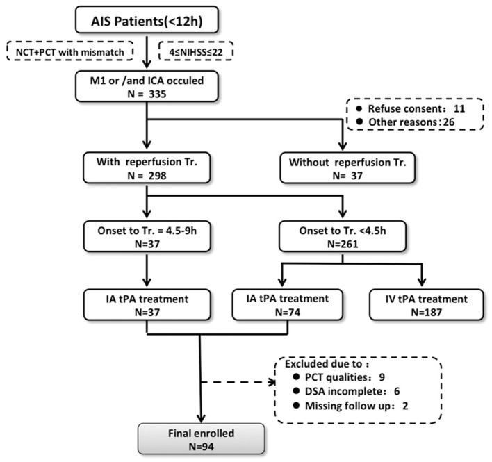
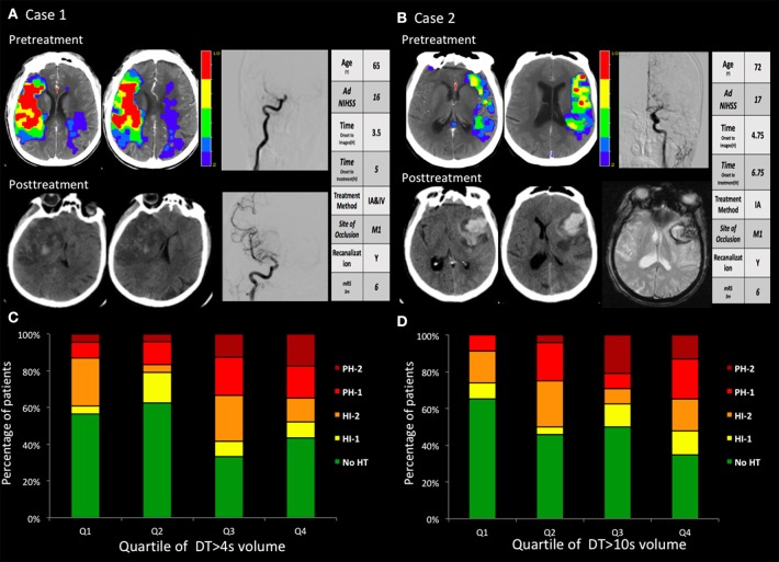
Cerebral hemorrhage is a serious potential complication of stroke revascularization, especially in patients receiving intra-arterial tissue-type plasminogen activator (tPA) therapy. We investigated the optimal pre-intervention delay time (DT) of computed tomography perfusion (CTP) measurement to predict cerebral parenchymal hematoma (PH) in acute ischemic stroke (AIS) patients after intra-arterial tissue plasminogen activator (tPA) treatment. The study population consisted of a series of patients with AIS who received intra-arterial tPA treatment and had CTP and follow-up computed tomography/magnetic resonance imaging (CT/MRI) to identify hemorrhagic transformation. The association of increasing DT thresholds (>2, >4, >6, >8, and >10 s) with PH was examined using receiver operating characteristic (ROC) analysis and logistic regression. Of 94 patients, 23 developed PH on follow-up imaging. Receiver operating characteristic analysis revealed that the greatest area under the curve for predicting PH occurred at DT > 4 s (area under the curve, 0.66). At this threshold of > 4 s, DT lesion volume ≥ 30.85 mL optimally predicted PH with 70% sensitivity and 59% specificity. DT > 4 s volume was independently predictive of PH in a multivariate logistic regression model ( < 0.05). DT > 4 s was the parameter most strongly associated with PH. The volume of moderate, not severe, hypo-perfusion on DT is more strongly associated and may allow better prediction of PH after intra-arterial tPA thrombolysis.

摘要

脑出血是卒中血管重建的一种严重潜在并发症,尤其是在接受动脉内组织型纤溶酶原激活剂(tPA)治疗的患者中。我们研究了计算机断层扫描灌注(CTP)测量的最佳干预前延迟时间(DT),以预测急性缺血性卒中(AIS)患者在接受动脉内组织纤溶酶原激活剂(tPA)治疗后的脑实质血肿(PH)。研究人群包括一系列接受动脉内tPA治疗并进行了CTP以及后续计算机断层扫描/磁共振成像(CT/MRI)以确定出血性转化的AIS患者。使用受试者操作特征(ROC)分析和逻辑回归检查了增加的DT阈值(>2、>4、>6、>8和>10秒)与PH的关联。在94例患者中,23例在随访成像时出现了PH。ROC分析显示,预测PH的曲线下面积在DT>4秒时最大(曲线下面积,0.66)。在这个>4秒的阈值下,DT病变体积≥30.85 mL对PH的预测最佳,灵敏度为70%,特异度为59%。在多变量逻辑回归模型中,DT>4秒的体积可独立预测PH(<0.05)。DT>4秒是与PH关联最密切的参数。DT上中度而非重度灌注不足的体积与之关联更强,可能有助于更好地预测动脉内tPA溶栓后的PH。

https://cdn.ncbi.nlm.nih.gov/pmc/blobs/5938/6110878/1a2fb1bf243e/fneur-09-00680-g0002.jpg
https://cdn.ncbi.nlm.nih.gov/pmc/blobs/5938/6110878/9fde91a6a205/fneur-09-00680-g0001.jpg
https://cdn.ncbi.nlm.nih.gov/pmc/blobs/5938/6110878/1a2fb1bf243e/fneur-09-00680-g0002.jpg
https://cdn.ncbi.nlm.nih.gov/pmc/blobs/5938/6110878/9fde91a6a205/fneur-09-00680-g0001.jpg
https://cdn.ncbi.nlm.nih.gov/pmc/blobs/5938/6110878/1a2fb1bf243e/fneur-09-00680-g0002.jpg

相似文献

1
Optimal Delay Time of CT Perfusion for Predicting Cerebral Parenchymal Hematoma After Intra-Arterial tPA Treatment.CT灌注预测动脉内注射组织型纤溶酶原激活剂治疗后脑实质血肿的最佳延迟时间
Front Neurol. 2018 Aug 21;9:680. doi: 10.3389/fneur.2018.00680. eCollection 2018.
2
Prediction of poststroke hemorrhagic transformation using computed tomography perfusion.基于 CT 灌注成像预测卒中后出血转化。
Stroke. 2013 Nov;44(11):3039-43. doi: 10.1161/STROKEAHA.113.002396. Epub 2013 Sep 3.
3
Trial design and reporting standards for intra-arterial cerebral thrombolysis for acute ischemic stroke.急性缺血性脑卒中动脉内脑溶栓的试验设计与报告标准。
Stroke. 2003 Aug;34(8):e109-37. doi: 10.1161/01.STR.0000082721.62796.09. Epub 2003 Jul 17.
4
Predictive accuracy of an ADC map for hemorrhagic transformation in acute ischemic stroke patients after successful recanalization with endovascular therapy.急性缺血性卒中患者经血管内治疗成功再通后,表观扩散系数(ADC)图对出血性转化的预测准确性。
Ann Transl Med. 2022 May;10(10):591. doi: 10.21037/atm-22-2255.
5
Reperfusion of very low cerebral blood volume lesion predicts parenchymal hematoma after endovascular therapy.极低脑血容量病变的再灌注可预测血管内治疗后的实质内血肿。
Stroke. 2015 May;46(5):1245-9. doi: 10.1161/STROKEAHA.114.008171. Epub 2015 Mar 31.
6
Two tales: hemorrhagic transformation but not parenchymal hemorrhage after thrombolysis is related to severity and duration of ischemia: MRI study of acute stroke patients treated with intravenous tissue plasminogen activator within 6 hours.两个故事:溶栓后出血性转化而非实质内出血与缺血的严重程度和持续时间相关:对6小时内接受静脉注射组织纤溶酶原激活剂治疗的急性卒中患者的MRI研究
Stroke. 2007 Feb;38(2):313-8. doi: 10.1161/01.STR.0000254565.51807.22. Epub 2007 Jan 4.
7
Hemorrhagic transformation of ischemic stroke: prediction with CT perfusion.缺血性卒中的出血性转化:CT灌注预测
Radiology. 2009 Mar;250(3):867-77. doi: 10.1148/radiol.2503080257.
8
High-permeability region size on perfusion CT predicts hemorrhagic transformation after intravenous thrombolysis in stroke.灌注CT上的高通透性区域大小可预测卒中静脉溶栓后的出血性转化。
PLoS One. 2017 Nov 28;12(11):e0188238. doi: 10.1371/journal.pone.0188238. eCollection 2017.
9
Validation of FLAIR hyperintense lesions as imaging biomarkers to predict the outcome of acute stroke after intra-arterial thrombolysis following intravenous tissue plasminogen activator.FLAIR 高信号病灶作为影像生物标志物预测静脉溶栓后动脉内溶栓治疗急性卒中的结局
Cerebrovasc Dis. 2013;35(5):461-8. doi: 10.1159/000350201. Epub 2013 May 31.
10
Mismatch of Low Perfusion and High Permeability Predicts Hemorrhagic Transformation Region in Acute Ischemic Stroke Patients Treated with Intra-arterial Thrombolysis.低灌注与高通透性不匹配可预测接受动脉内溶栓治疗的急性缺血性卒中患者的出血转化区域。
Sci Rep. 2016 Jun 15;6:27950. doi: 10.1038/srep27950.

引用本文的文献

1
Accuracy of Four Different CT Perfusion Thresholds for Ischemic Core Volume and Location Estimation Using IntelliSpace Portal.使用IntelliSpace Portal通过四种不同CT灌注阈值估计缺血核心体积和位置的准确性
J Cardiovasc Dev Dis. 2023 May 30;10(6):239. doi: 10.3390/jcdd10060239.
2
Perfusion CT for prediction of hemorrhagic transformation in acute ischemic stroke: a systematic review and meta-analysis.灌注 CT 预测急性缺血性脑卒中出血性转化:系统评价和荟萃分析。
Eur Radiol. 2019 Aug;29(8):4077-4087. doi: 10.1007/s00330-018-5936-7. Epub 2019 Jan 7.

本文引用的文献

1
Pretreatment blood-brain barrier disruption and post-endovascular intracranial hemorrhage.血管内介入颅内出血前的血脑屏障破坏
Neurology. 2016 Jul 19;87(3):263-9. doi: 10.1212/WNL.0000000000002862. Epub 2016 Jun 17.
2
Whole-Brain CT Perfusion to Quantify Acute Ischemic Penumbra and Core.全脑 CT 灌注成像定量评估急性缺血半暗带和核心区。
Radiology. 2016 Jun;279(3):876-87. doi: 10.1148/radiol.2015150319. Epub 2016 Jan 18.
3
The Heidelberg Bleeding Classification: Classification of Bleeding Events After Ischemic Stroke and Reperfusion Therapy.
海德堡出血分类:缺血性中风和再灌注治疗后出血事件的分类
Stroke. 2015 Oct;46(10):2981-6. doi: 10.1161/STROKEAHA.115.010049. Epub 2015 Sep 1.
4
Role of imaging in current acute ischemic stroke workflow for endovascular therapy.成像在当前急性缺血性卒中血管内治疗工作流程中的作用。
Stroke. 2015 Jun;46(6):1453-61. doi: 10.1161/STROKEAHA.115.009160. Epub 2015 May 5.
5
Perfusion computed tomography to assist decision making for stroke thrombolysis.灌注 CT 协助决策溶栓治疗脑卒中。
Brain. 2015 Jul;138(Pt 7):1919-31. doi: 10.1093/brain/awv071. Epub 2015 Mar 25.
6
Randomized assessment of rapid endovascular treatment of ischemic stroke.随机评估缺血性脑卒中的血管内治疗。
N Engl J Med. 2015 Mar 12;372(11):1019-30. doi: 10.1056/NEJMoa1414905. Epub 2015 Feb 11.
7
Endovascular therapy for ischemic stroke with perfusion-imaging selection.血管内治疗缺血性卒中的灌注成像选择。
N Engl J Med. 2015 Mar 12;372(11):1009-18. doi: 10.1056/NEJMoa1414792. Epub 2015 Feb 11.
8
A randomized trial of intraarterial treatment for acute ischemic stroke.急性缺血性脑卒中的动脉内治疗随机试验。
N Engl J Med. 2015 Jan 1;372(1):11-20. doi: 10.1056/NEJMoa1411587. Epub 2014 Dec 17.
9
Pretreatment blood-brain barrier damage and post-treatment intracranial hemorrhage in patients receiving intravenous tissue-type plasminogen activator.接受静脉注射组织型纤溶酶原激活物的患者的预处理血脑屏障损伤和治疗后颅内出血。
Stroke. 2014 Jul;45(7):2030-5. doi: 10.1161/STROKEAHA.114.005249. Epub 2014 May 15.
10
Prediction of poststroke hemorrhagic transformation using computed tomography perfusion.基于 CT 灌注成像预测卒中后出血转化。
Stroke. 2013 Nov;44(11):3039-43. doi: 10.1161/STROKEAHA.113.002396. Epub 2013 Sep 3.