Rodriguez Léa, Mdzomba Julius Baya, Joly Sandrine, Boudreau-Laprise Mélissa, Planel Emmanuel, Pernet Vincent
CUO-Recherche, Centre de Recherche du CHU de Québec, Quebec, QC, Canada.
Département d'ophtalmologie, Faculté de Médecine, Université Laval, Quebec, QC, Canada.
Front Mol Neurosci. 2018 Aug 24;11:293. doi: 10.3389/fnmol.2018.00293. eCollection 2018.
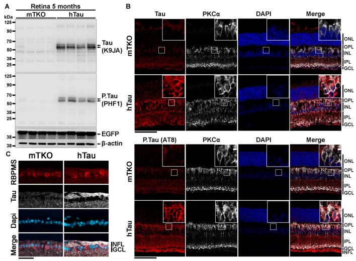
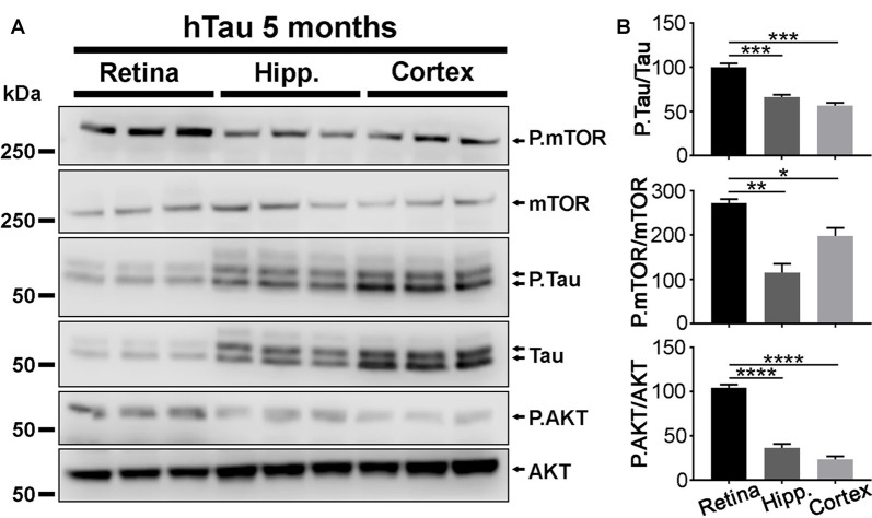
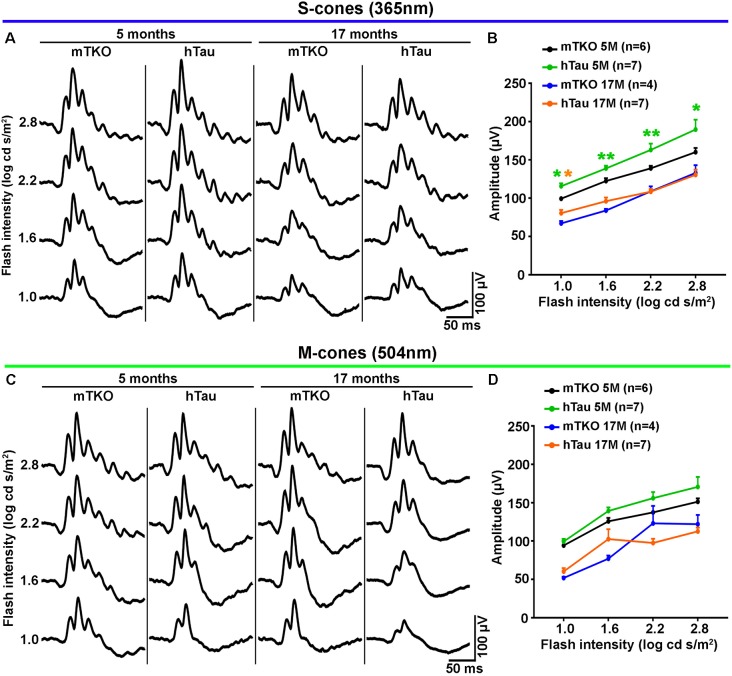
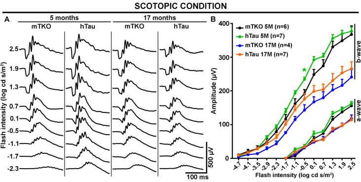
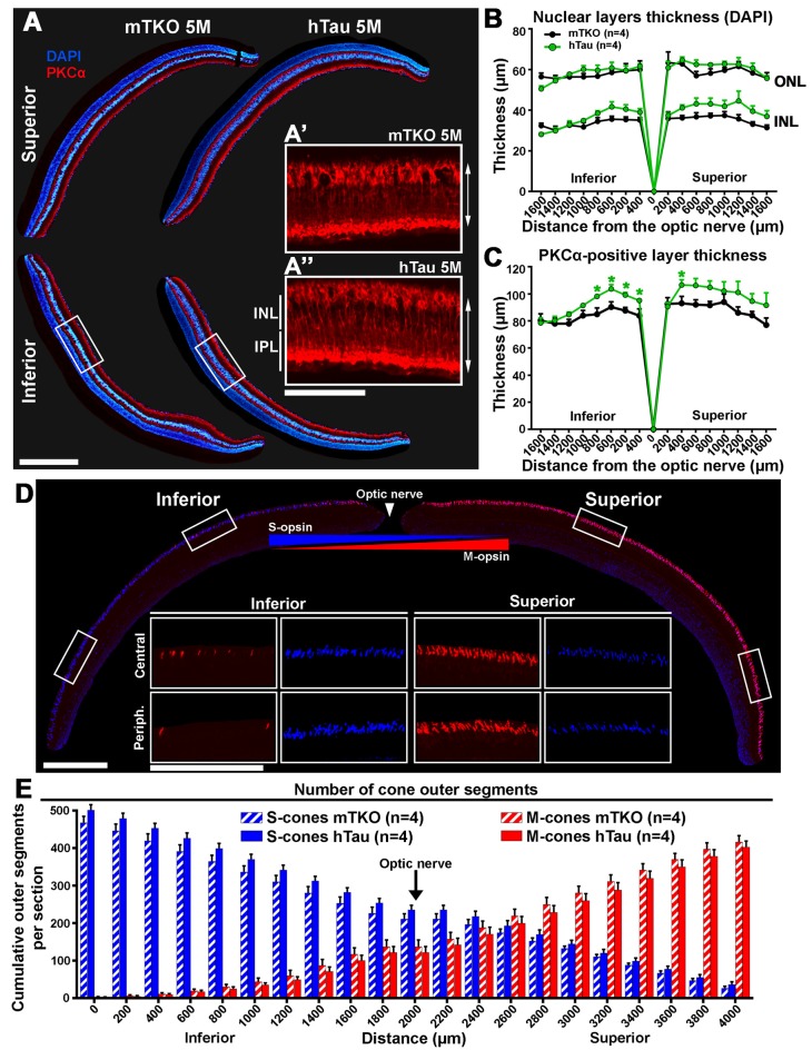
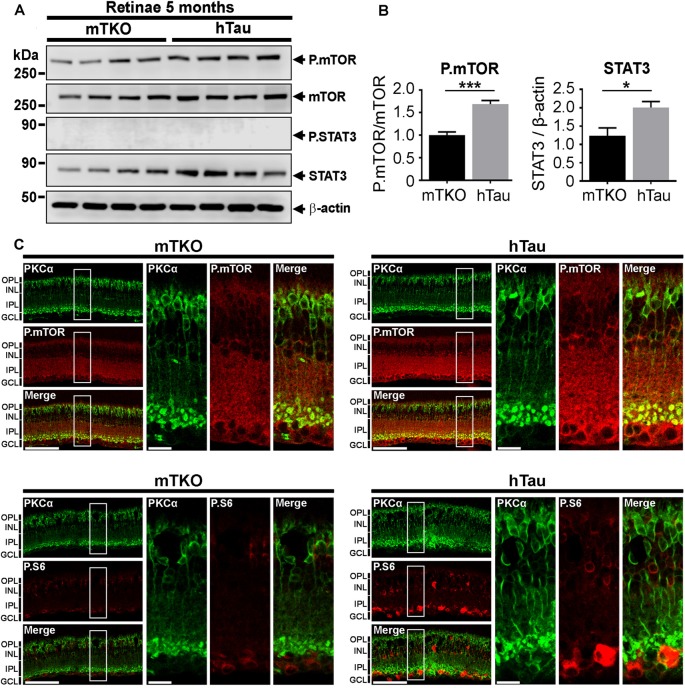
The implication of the microtubule-associated protein (MAP) Tau in the ocular manifestations of Alzheimer's disease (AD) is elusive due to the lack of relevant animal model. However, signs of AD have been reported in the brain of transgenic mice expressing human Tau (hTau). To assess whether hTau is sufficient to induce AD pathogenesis in the retina as well, in the present study, we compared the retinal structure and function of KO mice deprived of Tau (mTKO) with those of transgenic mice expressing hTau. Our results revealed that hTau is particularly abundant in the inner nuclear layer (INL) cells of the retina. By electroretinogram (ERG) recording, light-induced retinal cell activation was not altered in hTau compared with mTKO littermates. Surprisingly, the ERG response mediated by cone photoreceptor stimulation was even stronger in hTau than in mTKO retinae. Immunofluorescent analysis of retinal sections allowed us to observe thicker inner retina in hTau than in mTKO eyes. By Western Blotting (WB), the upregulation of mTOR that was found in hTau mice may underlie retinal structure and function increases. Taken together, our results not only indicate that hTau expression is not toxic for retinal cells but they also suggest that it may play a positive role in visual physiology. The use of hTau may be envisaged to improve visual recovery in ocular diseases affecting the retinal function such as glaucoma or diabetic retinopathy.
由于缺乏相关动物模型,微管相关蛋白(MAP)Tau在阿尔茨海默病(AD)眼部表现中的作用尚不清楚。然而,在表达人Tau(hTau)的转基因小鼠大脑中已报道有AD迹象。为了评估hTau是否也足以在视网膜中诱导AD发病机制,在本研究中,我们比较了缺乏Tau的敲除小鼠(mTKO)与表达hTau的转基因小鼠的视网膜结构和功能。我们的结果显示,hTau在视网膜内核层(INL)细胞中特别丰富。通过视网膜电图(ERG)记录,与同窝mTKO小鼠相比,hTau小鼠中光诱导的视网膜细胞激活没有改变。令人惊讶的是,在hTau小鼠中,由视锥光感受器刺激介导的ERG反应甚至比mTKO视网膜更强。对视网膜切片的免疫荧光分析使我们观察到,hTau小鼠的视网膜内层比mTKO小鼠的更厚。通过蛋白质免疫印迹法(WB),在hTau小鼠中发现的mTOR上调可能是视网膜结构和功能增加的基础。综上所述,我们的结果不仅表明hTau表达对视网膜细胞无毒,还表明它可能在视觉生理学中发挥积极作用。可以设想使用hTau来改善影响视网膜功能的眼部疾病(如青光眼或糖尿病性视网膜病变)中的视觉恢复。