,.
Invest Ophthalmol Vis Sci. 2020 Jun 3;61(6):12. doi: 10.1167/iovs.61.6.12.
Synucleinopathies such as multiple system atrophy (MSA) and Parkinson's disease are associated with a variety of visual symptoms. Functional and morphological retinal aberrations are therefore supposed to be valuable biomarkers for these neurodegenerative diseases. This study examined the retinal morphology and functionality resulting from human α-synuclein (α-Syn) overexpression in the transgenic Plp-α-Syn mouse model.
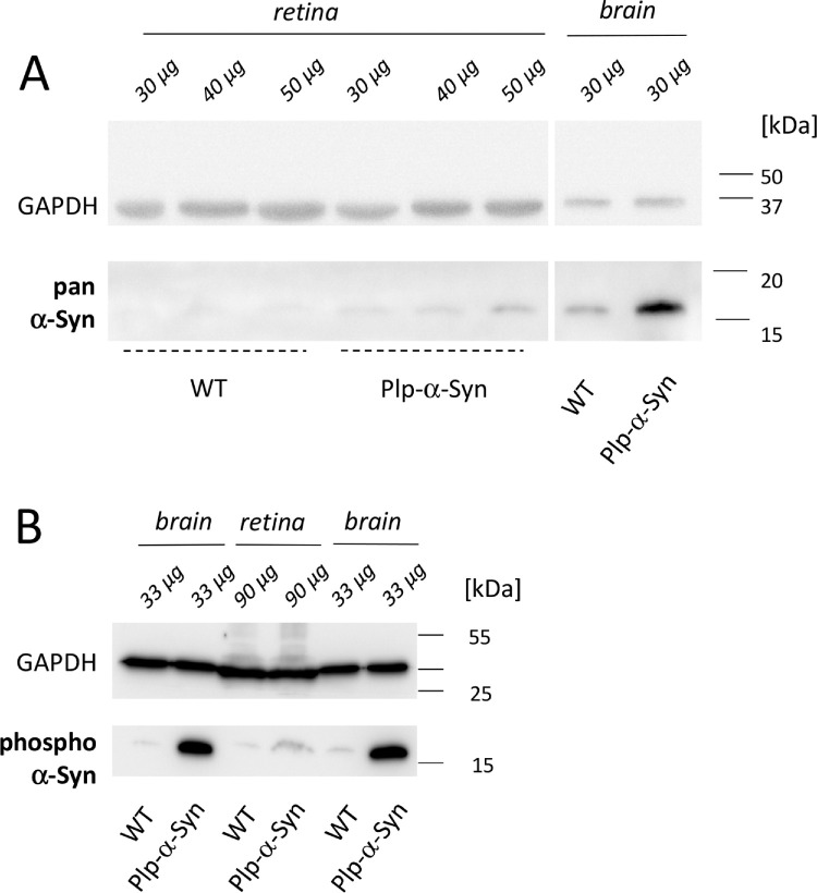
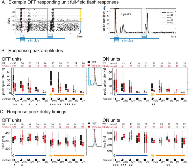
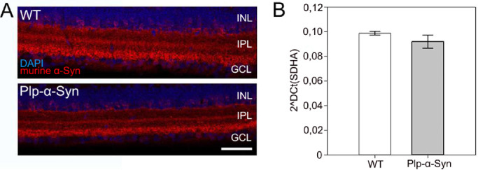
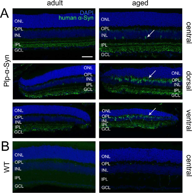
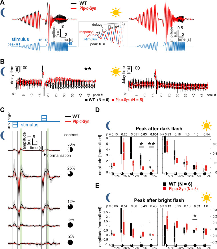
Immunohistochemistry on retinal sections and whole-mounts was performed on 8- to 11-week-old and 12-month-old Plp-α-Syn mice and C57BL/6N controls. Quantitative RT-PCR experiments were performed to study the expression of endogenous and human α-Syn and tyrosine hydroxylase (TH). We confirmed the presence of human α-Syn in the retina in western blot analyses. Multi-electrode array (MEA) analyses from light-stimulated whole-mounted retinas were used to investigate their functionality.
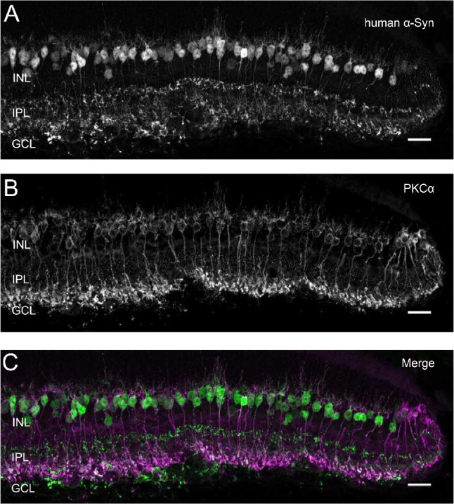
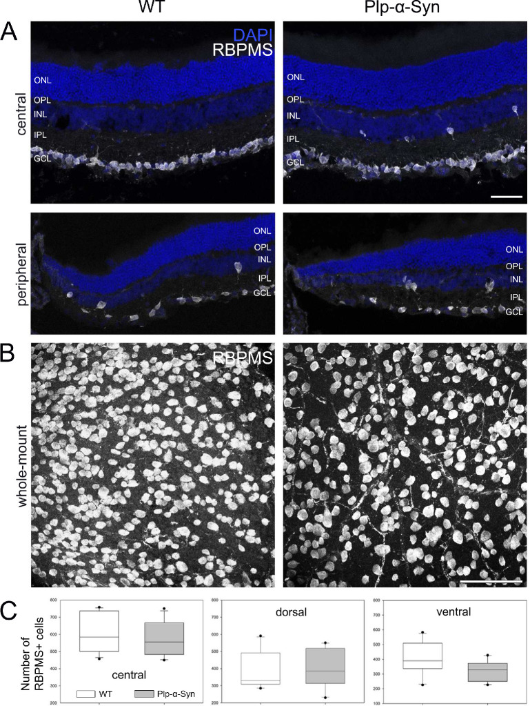
Biochemical and immunohistochemical analyses showed human α-Syn in the retina of Plp-α-Syn mice. We found distinct staining in different retinal cell layers, most abundantly in rod bipolar cells of the peripheral retina. In the periphery, we also observed a trend toward a decline in the number of retinal ganglion cells. The number of TH+ neurons was unaffected in this human α-Syn overexpression model. MEA recordings showed that Plp-α-Syn retinas were functional but exhibited mild alterations in dim light conditions.
Together, these findings implicate an impairment of retinal neurons in the Plp-α-Syn mouse. The phenotype partly relates to retinal deficits reported in MSA patients. We further propose the suitability of the Plp-α-Syn retina as a biological model to study synuclein-mediated mechanisms.
多种系统萎缩症(MSA)和帕金森病等突触核蛋白病与多种视觉症状有关。因此,功能和形态视网膜异常被认为是这些神经退行性疾病的有价值的生物标志物。本研究检查了人类α-突触核蛋白(α-Syn)在转基因 Plp-α-Syn 小鼠模型中过度表达导致的视网膜形态和功能。
对 8-11 周龄和 12 月龄的 Plp-α-Syn 小鼠和 C57BL/6N 对照进行视网膜切片和全视网膜免疫组织化学染色。进行定量 RT-PCR 实验以研究内源性和人类 α-Syn 和酪氨酸羟化酶(TH)的表达。我们通过 Western blot 分析证实了视网膜中存在人类 α-Syn。使用光刺激全视网膜多电极阵列(MEA)分析来研究其功能。
生化和免疫组织化学分析显示 Plp-α-Syn 小鼠的视网膜中有人类 α-Syn。我们在不同的视网膜细胞层中发现了明显的染色,在周边视网膜的杆状双极细胞中最为丰富。在周边,我们还观察到视网膜神经节细胞数量减少的趋势。在这个人类 α-Syn 过度表达模型中,TH+神经元的数量没有受到影响。MEA 记录显示 Plp-α-Syn 视网膜具有功能,但在弱光条件下表现出轻微改变。
这些发现表明 Plp-α-Syn 小鼠的视网膜神经元受损。该表型部分与 MSA 患者报告的视网膜缺陷有关。我们进一步提出 Plp-α-Syn 视网膜作为研究突触核蛋白介导机制的生物模型的适用性。