Department of Anatomy and Neurobiology, Graduate School of Medical Sciences, Kumamoto University, 1-1-1 Honjo, Chuo-ku, Kumamoto, 860-8556, Japan.
Department of Pharmacology, Kurume University, Kurume, 830-0111, Japan.
Brain Struct Funct. 2018 Dec;223(9):4275-4291. doi: 10.1007/s00429-018-1749-3. Epub 2018 Sep 10.
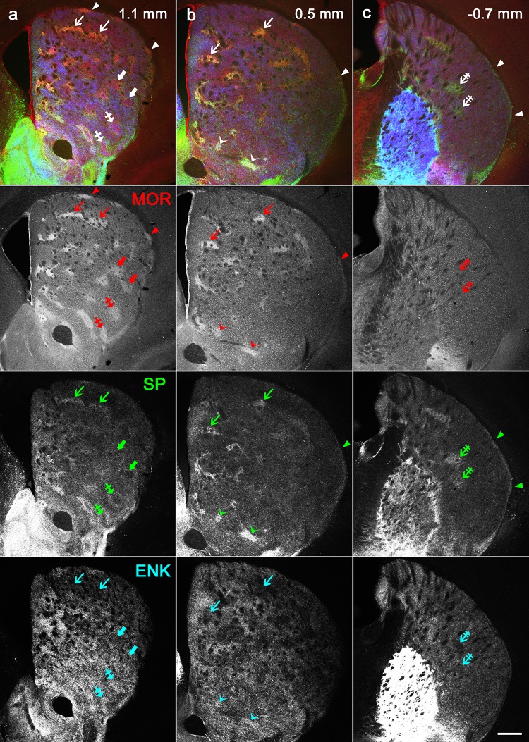
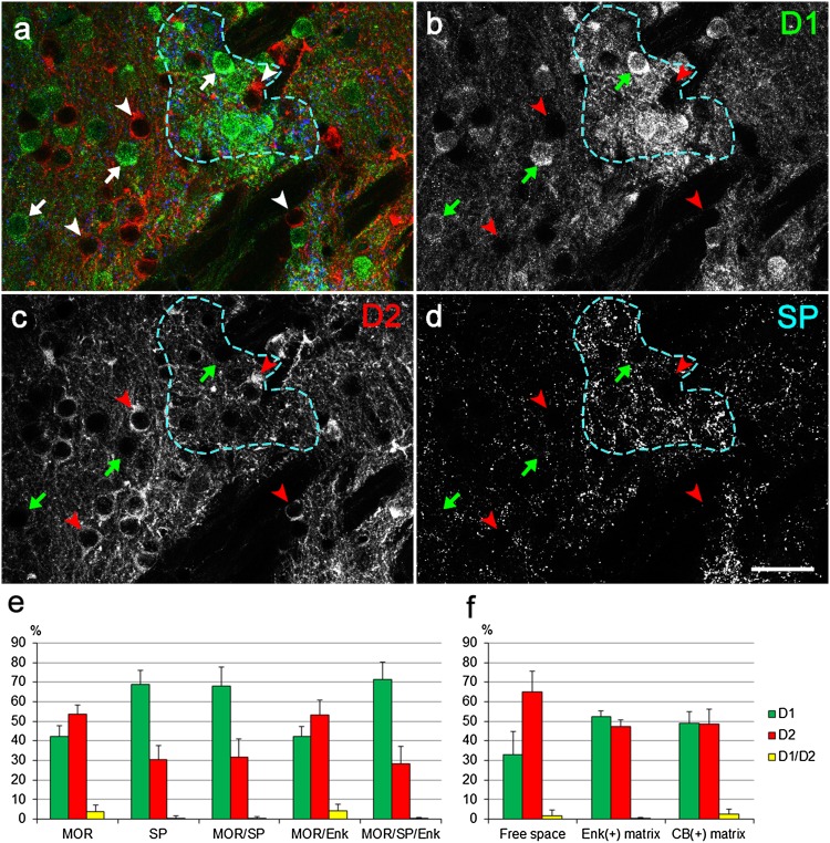
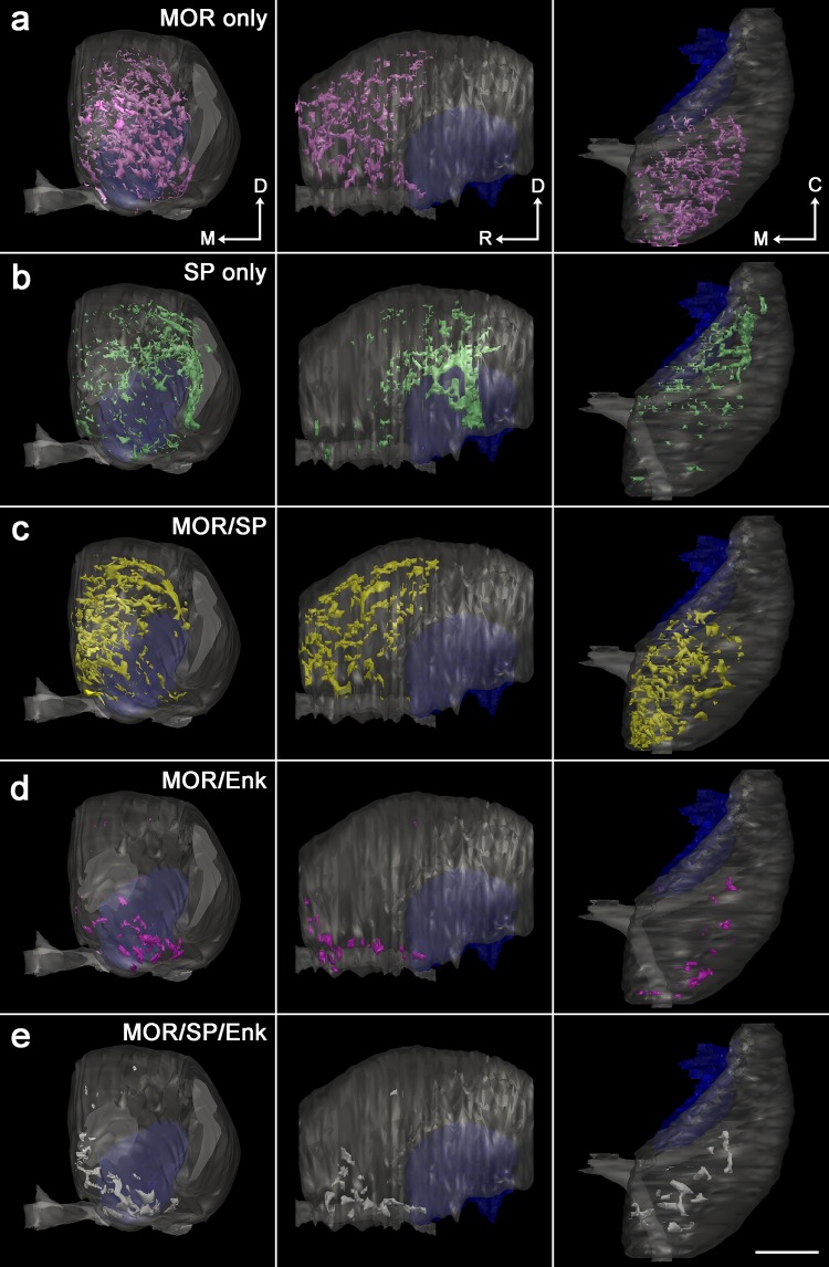
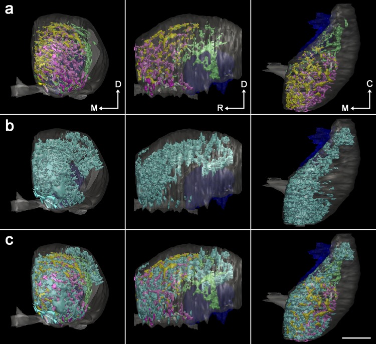
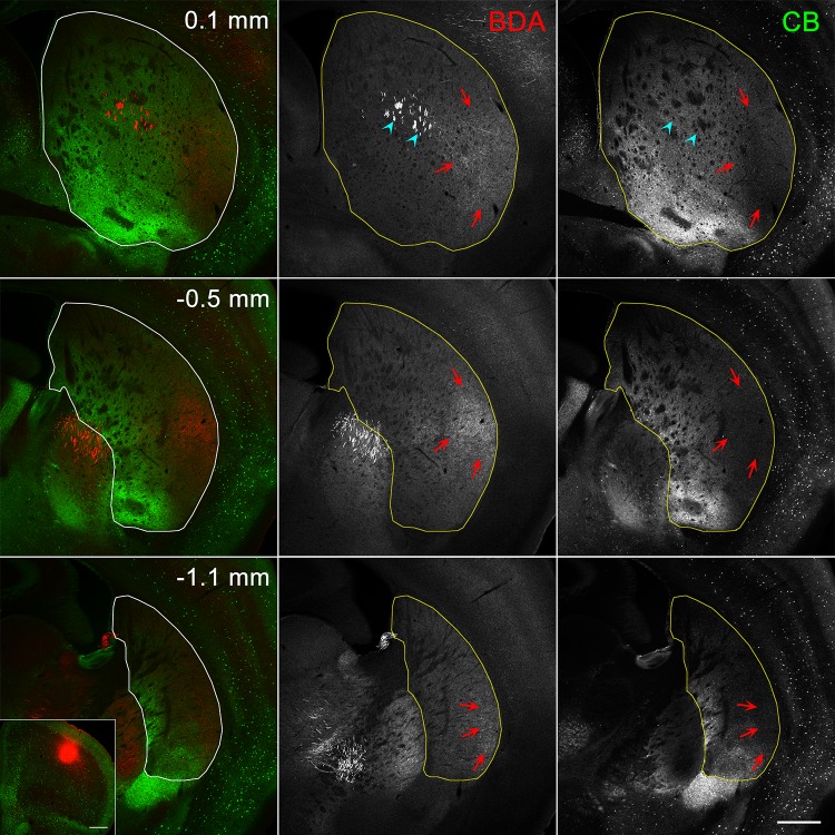
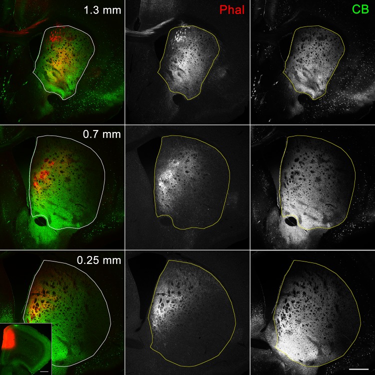
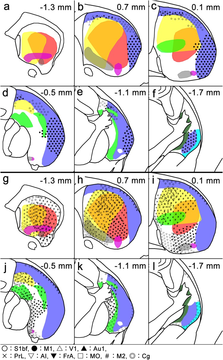
The striatum is critically involved in execution of appropriate behaviors, but its internal structures remain unmapped due to its unique structural organization, leading to ambiguity when interpreting heterogeneous properties of striatal neurons that differ by location. We focused on site-specific diversity of striosomes/matrix compartmentalization to draw the striatum map. Five types of striosomes were discriminated according to diverse immunoreactivities for the µ-opioid receptor, substance P (SP) and enkephalin, and each type occupied a particular domain inside the striatum. Furthermore, there was an additional domain lacking striosomes. This striosome-free space was located at the dorsolateral region and received afferents preferentially from the primary motor and sensory cortices, whereas the striosome-rich part received afferents from associational/limbic cortices, with topography inside both innervations. The proportion of dopamine D1 receptor-expressing, presumptive striatonigral neurons was approximately 70% in SP-positive striosomes, 40% in SP-deficient striosomes, 30% in the striosome-free space, and 50% in the matrix. In contrast, the proportion of D2 receptor-expressing, presumptive striatopallidal neurons was complementary to that of D1 receptor-expressing cells, indicating a close relationship between the map and the direct and indirect parallel circuitry. Finally, the most caudal part of the striatum lacked compartmentalization and consisted of three lamina characterized by intense and mutually exclusive immunoreactivities for SP and enkephalin. This tri-laminar part also received specific afferents from the cortex. The newly obtained map will facilitate broad fields of research in the basal ganglia with higher resolution of the three-dimensional anatomy of the striatum.
纹状体在执行适当的行为方面起着至关重要的作用,但由于其独特的结构组织,其内部结构仍未被绘制出来,这导致在解释纹状体神经元的异质特性时存在歧义,而这些特性因位置而异。我们专注于纹状体/基质分区的特定部位多样性,以绘制纹状体图谱。根据µ-阿片受体、P 物质 (SP) 和脑啡肽的不同免疫反应性,区分了五种类型的纹状体,每种类型都占据了纹状体内部的特定区域。此外,还有一个没有纹状体的额外区域。这个没有纹状体的空间位于背外侧区域,优先接收来自初级运动和感觉皮层的传入,而富含纹状体的部分则接收来自联合/边缘皮层的传入,在这两种传入中都有拓扑结构。多巴胺 D1 受体表达的、假定的纹状体苍白球神经元在 SP 阳性纹状体中的比例约为 70%,在 SP 缺乏的纹状体中的比例约为 40%,在没有纹状体的空间中的比例约为 30%,在基质中的比例约为 50%。相比之下,D2 受体表达的、假定的纹状体黑质神经元的比例与 D1 受体表达细胞互补,表明图谱与直接和间接平行电路之间存在密切关系。最后,纹状体的最尾端部分缺乏分区,由三个层组成,这些层的特征是 SP 和脑啡肽的免疫反应性强烈且相互排斥。这个三层部分还接收来自皮层的特定传入。新获得的图谱将促进基底神经节更广泛的研究领域,提高纹状体三维解剖结构的分辨率。