a Department of Surgical Sciences , Section of Otolaryngology, Uppsala University Hospital , Uppsala , Sweden.
b Department of Otolaryngology , Peking University Shenzhen Hospital , P.R. China.
Ups J Med Sci. 2018 Sep;123(3):131-142. doi: 10.1080/03009734.2018.1492654. Epub 2018 Sep 11.
The Uppsala collection of human temporal bones and molds is a unique resource for education and international research collaboration. Micro-computerized tomography (micro-CT) and synchrotron imaging are used to investigate the complex anatomy of the inner ear. Impaired microcirculation is etiologically linked to various inner ear disorders, and recent developments in inner ear surgery promote examination of the vascular system. Here, for the first time, we present three-dimensional (3D) data from investigations of the major vascular pathways and corresponding bone channels.
We used the archival Uppsala collection of temporal bones and molds consisting of 324 inner ear casts and 113 macerated temporal bones. Micro-CT was used to investigate vascular bone channels, and 26 fresh human temporal bones underwent synchrotron radiation phase contrast imaging (SR-PCI). Data were processed by volume-rendering software to create 3D reconstructions allowing orthogonal sectioning, cropping, and soft tissue analyses.
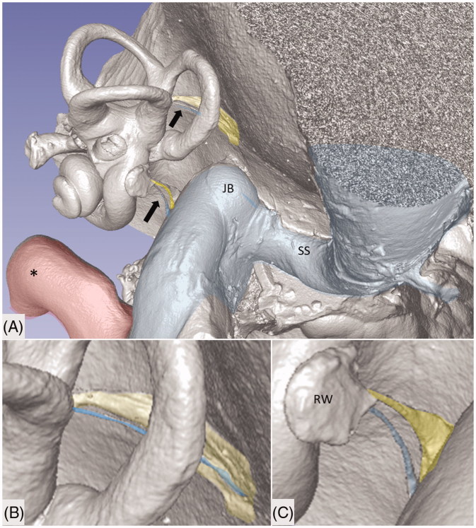
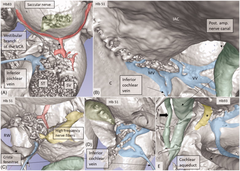
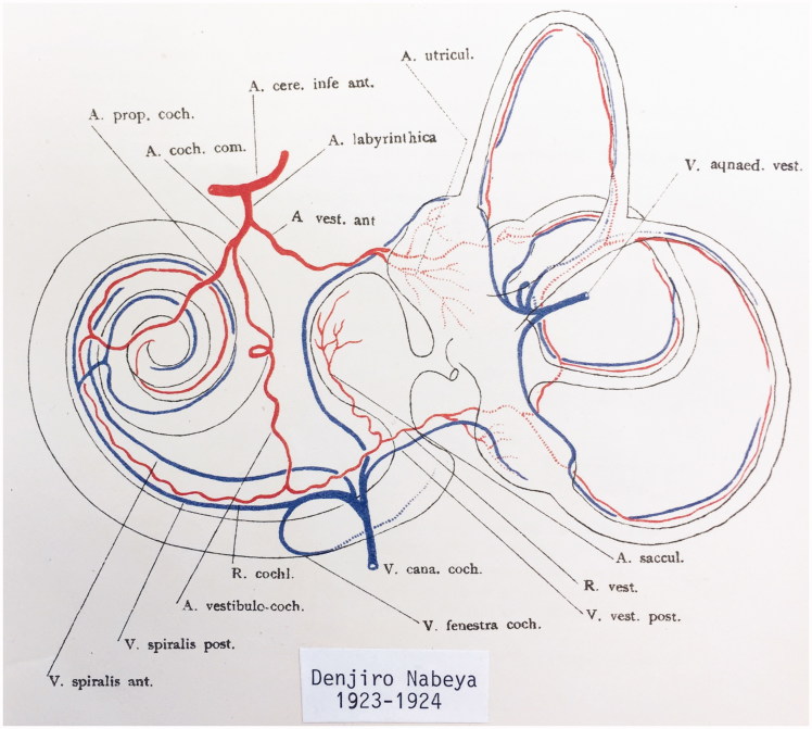
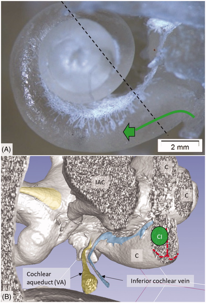
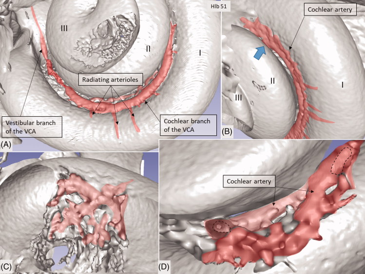
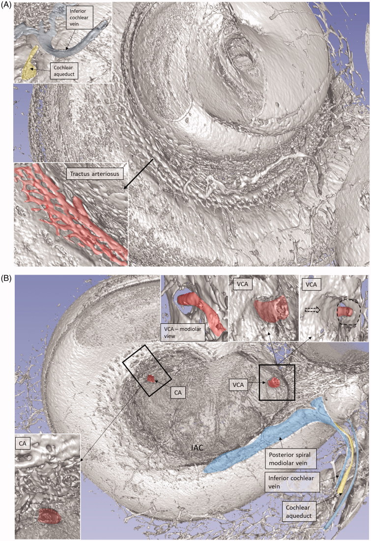
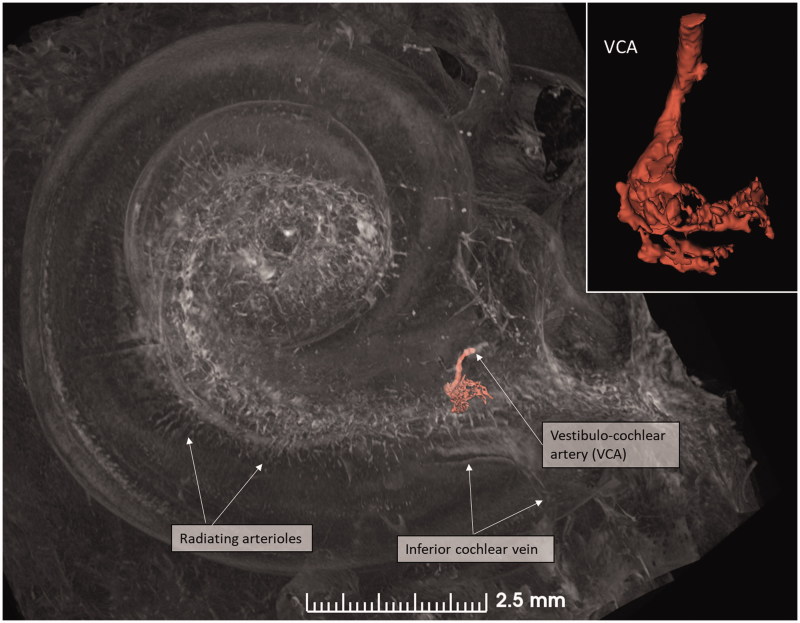
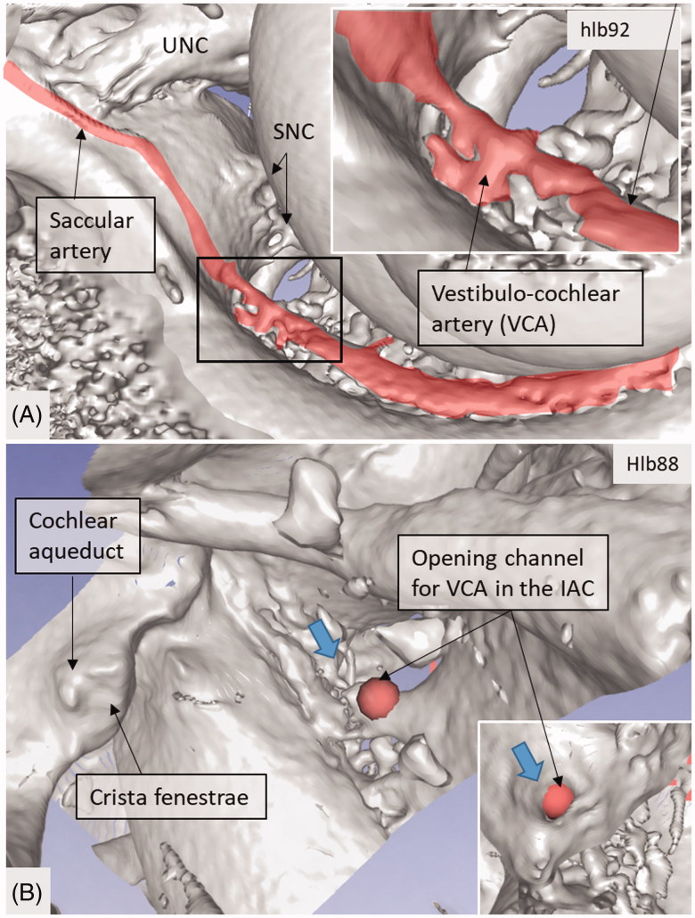
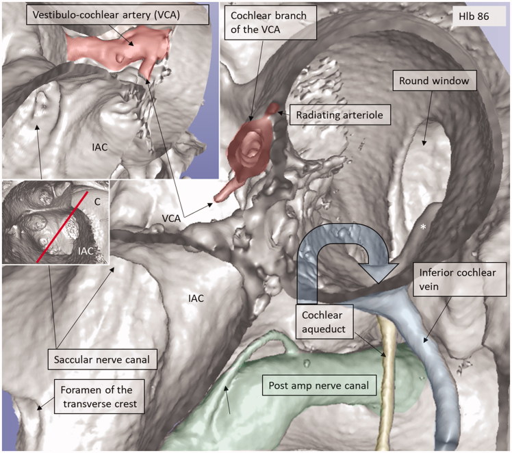
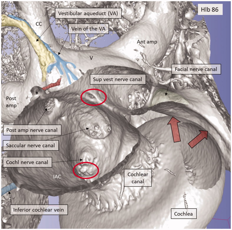
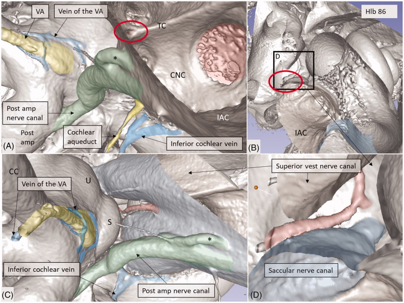
Micro-CT with 3D rendering was superior in reproducing the anatomy of the vascular bone channels, while SR-PCI replicated soft tissues. Arterial bone channels were traced from scala vestibuli (SV) arterioles to the fundus, cochlea, and vestibular apparatus. Drainage routes along the aqueducts were examined.
Human inner ear vessels are difficult to study due to the adjoining hard bone. Micro-CT and SR-PCI with 3D reconstructions revealed large portions of the micro-vascular system in un-decalcified specimens. The results increase our understanding of the organization of the vascular system in humans and how altered microcirculation may relate to inner ear disorders. The findings may also have surgical implications.
乌普萨拉人类颞骨和模具收藏是教育和国际研究合作的独特资源。微计算机断层扫描(micro-CT)和同步辐射成像用于研究内耳的复杂解剖结构。微循环受损与各种内耳疾病有关,内耳手术的最新进展促进了对血管系统的检查。在这里,我们首次展示了对主要血管途径和相应骨道进行三维(3D)研究的结果。
我们使用了由 324 个内耳铸模和 113 个脱钙颞骨组成的乌普萨拉存档颞骨和模具收藏。微 CT 用于研究血管骨通道,26 个新鲜人类颞骨接受了同步辐射相衬成像(SR-PCI)。数据通过体绘制软件进行处理,以创建允许正交切片、裁剪和软组织分析的 3D 重建。
3D 渲染的微 CT 在再现血管骨通道的解剖结构方面更具优势,而 SR-PCI 则复制了软组织。动脉骨通道从前庭小管(SV)小动脉追踪到基底、耳蜗和前庭器官。还检查了沿着导水管的引流途径。
由于毗邻的硬骨,人类内耳血管难以研究。微 CT 和带有 3D 重建的 SR-PCI 揭示了未脱钙标本中小血管系统的大部分。研究结果增加了我们对内耳血管系统在人类中的组织和微循环改变如何与内耳疾病相关的理解。这些发现也可能具有手术意义。