Tian Jun, Yan Yaping, Xi Wang, Zhou Rui, Lou Huifang, Duan Shumin, Chen Jiang Fan, Zhang Baorong
Second Affiliated Hospital, Zhejiang University School of Medicine, Hangzhou, China.
Department of Neurobiology, School of Medicine, Zhejiang University, Hangzhou, China.
Front Behav Neurosci. 2018 Aug 27;12:185. doi: 10.3389/fnbeh.2018.00185. eCollection 2018.
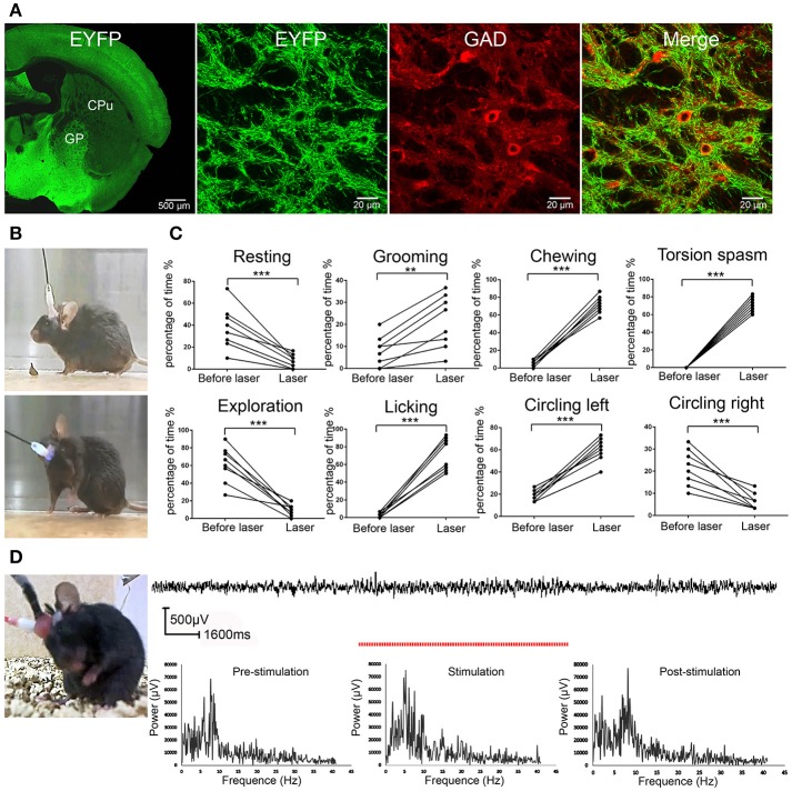
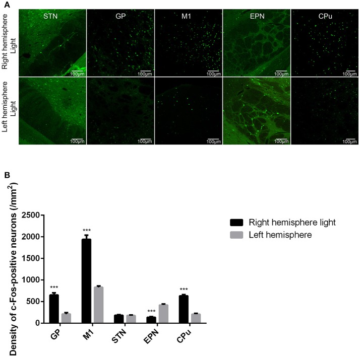
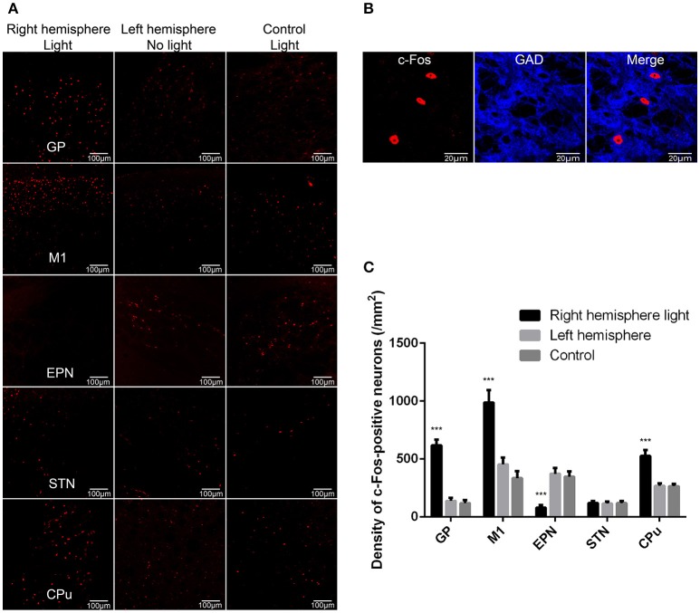
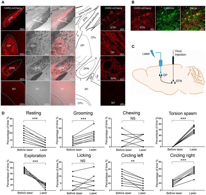
The globus pallidus (GP) is emerging as a critical locus of basal ganglia control of motor activity, but the exact role of GABAergic GP neurons remain to be defined. By targeted expression of channelrhodopsin 2 (ChR2) in GABAergic neurons using the VGAT-ChR2-EYFP transgenic mice, we showed that optogenetic stimulation of GABAergic neurons in the right GP produced hyperkinesia. Optogenetic stimulation of GABAergic GP neurons increased c-Fos-positive cells in GP, M1 cortex, and caudate-putamen (CPu), and decreased c-Fos-positive cells in entopeduncular nucleus (EPN), compared to the contralateral hemisphere. In agreement with the canonical basal ganglia model. Furthermore, we delivered AAV-CaMKIIα-ChR2-mCherry virus to the excitatory neurons of the subthalamic nucleus (STN) and selectively stimulated glutamatergic afferent fibers from the STN onto the GP. This optogenetic stimulation produced abnormal movements, similar to the behaviors that observed in the VGAT-ChR2-EYFP transgenic mice. Meanwhile, we found that the c-Fos expression pattern in the GP, M1, STN, EPN, and CPu produced by optogenetic activation of glutamatergic afferent fibers from the STN in GP was similar to the c-Fos expression pattern in the VGAT-ChR2-EYFP transgenic mice. Taken together, our results suggest that excess GP GABAergic neurons activity could be the neural substrate of abnormal involuntary movements in hyperkinetic movement disorders. The neural circuitry underlying the abnormal involuntary movements is associated with excessive GP, M1, CPu activity, and reduced EPN activity. Inhibition of GP GABAergic neurons represents new treatment targets for hyperkinetic movement disorder.
苍白球(GP)正逐渐成为基底神经节控制运动活动的关键部位,但GABA能苍白球神经元的确切作用仍有待确定。通过使用VGAT-ChR2-EYFP转基因小鼠在GABA能神经元中靶向表达通道视紫红质2(ChR2),我们发现对右侧苍白球中的GABA能神经元进行光遗传学刺激会产生运动亢进。与对侧半球相比,对苍白球GABA能神经元进行光遗传学刺激会增加苍白球、初级运动皮层(M1)和尾状核-壳核(CPu)中c-Fos阳性细胞的数量,并减少内苍白球核(EPN)中c-Fos阳性细胞的数量。这与经典的基底神经节模型一致。此外,我们将腺相关病毒-CaMKIIα-ChR2-mCherry病毒注射到丘脑底核(STN)的兴奋性神经元中,并选择性地刺激从STN到苍白球的谷氨酸能传入纤维。这种光遗传学刺激产生了异常运动,类似于在VGAT-ChR2-EYFP转基因小鼠中观察到的行为。同时,我们发现由苍白球中STN的谷氨酸能传入纤维的光遗传学激活所产生的苍白球、M1、STN、EPN和CPu中的c-Fos表达模式与VGAT-ChR2-EYFP转基因小鼠中的c-Fos表达模式相似。综上所述,我们的结果表明,苍白球GABA能神经元活动过多可能是运动亢进性运动障碍中异常不自主运动的神经基础。异常不自主运动背后的神经回路与苍白球、M1、CPu活动过多以及EPN活动减少有关。抑制苍白球GABA能神经元代表了运动亢进性运动障碍的新治疗靶点。