Department of Medicinal Chemistry, Uppsala University, Sweden.
Department of Protein Science, School of Engineering Sciences in Chemistry, Biotechnology and Health, KTH Royal Institute of Technology, Sweden.
Theranostics. 2018 Aug 7;8(16):4462-4476. doi: 10.7150/thno.24395. eCollection 2018.
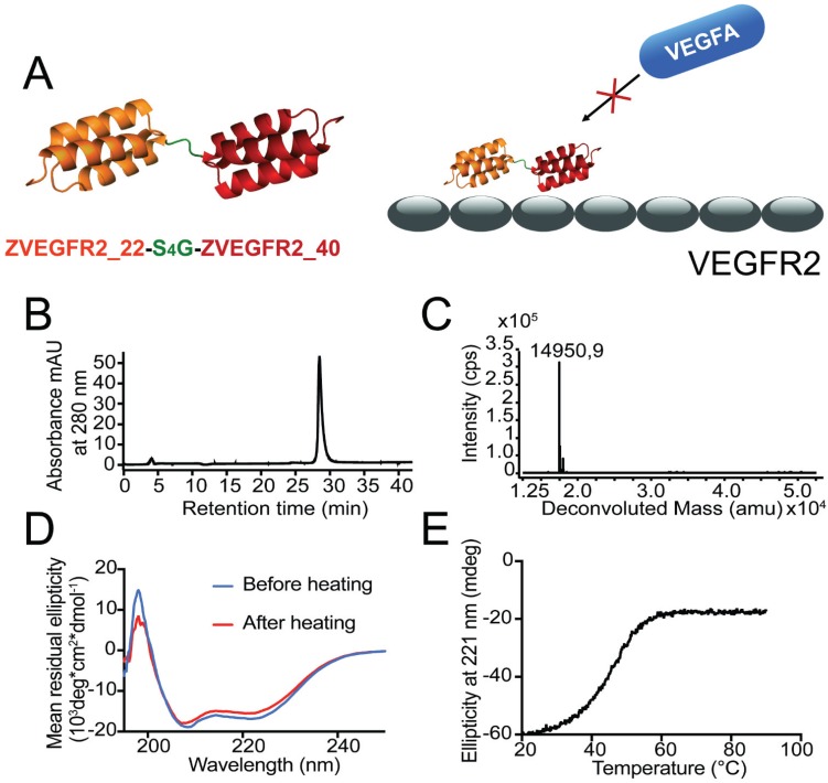
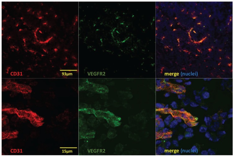
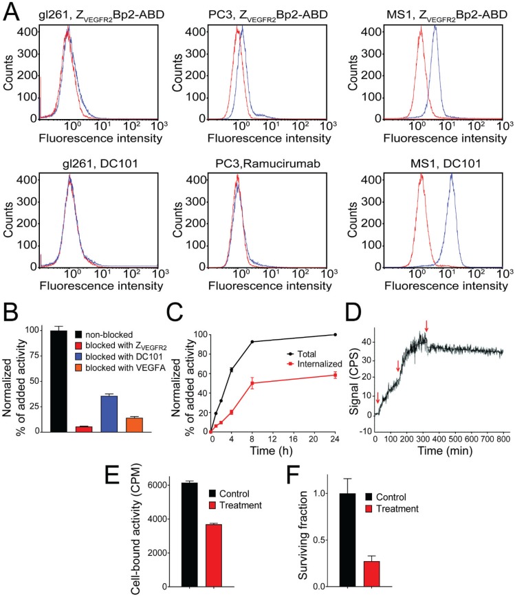
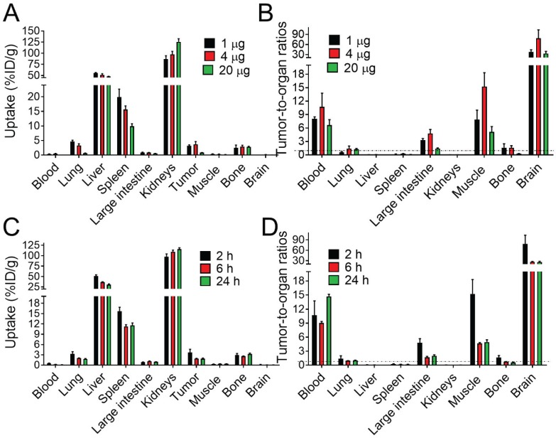
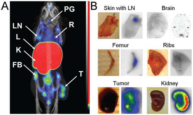
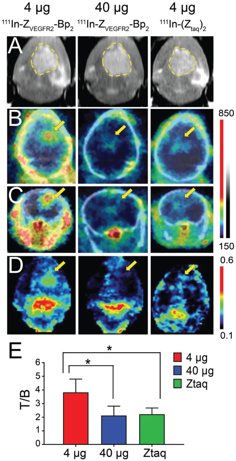
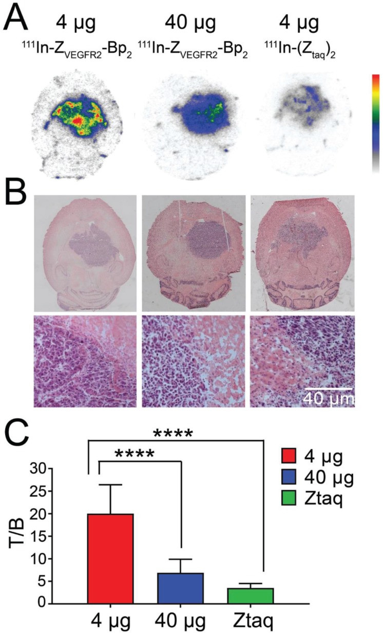
Vascular endothelial growth factor receptor-2 (VEGFR2) is a key mediator of angiogenesis and therefore a promising therapeutic target in malignancies including glioblastoma multiforme (GBM). Molecular imaging of VEGFR2 expression may enable patient stratification for antiangiogenic therapy. The goal of the current study was to evaluate the capacity of the novel anti-VEGFR2 biparatopic affibody conjugate (Z-Bp) for in vivo visualization of VEGFR2 expression in GBM. Z-Bp coupled to a NODAGA chelator was generated and radiolabeled with indium-111. The VEGFR2-expressing murine endothelial cell line MS1 was used to evaluate in vitro binding specificity and affinity, cellular processing and targeting specificity in mice. Further tumor targeting was studied in vivo in GL261 glioblastoma orthotopic tumors. Experimental imaging was performed. [In]In-NODAGA-Z-Bp bound specifically to VEGFR2 (K=33±18 pM). VEGFR2-mediated accumulation was observed in liver, spleen and lungs. The tumor-to-organ ratios 2 h post injection for mice bearing MS1 tumors were approximately 11 for blood, 15 for muscles and 78 for brain. Intracranial GL261 glioblastoma was visualized using SPECT/CT. The activity uptake in tumors was significantly higher than in normal brain tissue. The tumor-to-cerebellum ratios after injection of 4 µg [In]In-NODAGA-Z-Bp were significantly higher than the ratios observed for the 40 µg injected dose and for the non-VEGFR2 binding size-matched conjugate, demonstrating target specificity. Microautoradiography of cryosectioned CNS tissue was in good agreement with the SPECT/CT images. The anti-VEGFR2 affibody conjugate [In]In-NODAGA-Z-Bp specifically targeted VEGFR2 in vivo and visualized its expression in a murine GBM orthotopic model. Tumor-to-blood ratios for [In]In-NODAGA-Z-Bp were higher compared to other VEGFR2 imaging probes. [In]In-NODAGA-Z-Bp appears to be a promising probe for in vivo noninvasive visualization of tumor angiogenesis in glioblastoma.
血管内皮生长因子受体-2(VEGFR2)是血管生成的关键介质,因此是包括多形性胶质母细胞瘤(GBM)在内的恶性肿瘤的有前途的治疗靶点。VEGFR2 表达的分子成像可以实现抗血管生成治疗的患者分层。本研究的目的是评估新型抗-VEGFR2 双价亲和体结合物(Z-Bp)在体内可视化 GBM 中 VEGFR2 表达的能力。 生成与 NODAGA 螯合剂偶联的 Z-Bp,并与铟-111 标记。使用表达 VEGFR2 的鼠内皮细胞系 MS1 评估体外结合特异性和亲和力、细胞处理和在小鼠中的靶向特异性。进一步在 GL261 胶质母细胞瘤原位肿瘤中进行体内肿瘤靶向研究。进行了实验成像。 [In]In-NODAGA-Z-Bp 特异性结合 VEGFR2(K=33±18 pM)。在肝脏、脾脏和肺部观察到 VEGFR2 介导的积累。携带 MS1 肿瘤的小鼠注射后 2 小时的肿瘤与器官比值约为血液的 11,肌肉的 15,大脑的 78。使用 SPECT/CT 可视化颅内 GL261 胶质母细胞瘤。肿瘤的摄取活性明显高于正常脑组织。注射 4 µg [In]In-NODAGA-Z-Bp 后肿瘤与小脑的比值明显高于注射 40 µg 剂量和非 VEGFR2 结合大小匹配的缀合物观察到的比值,表明了靶向特异性。冷冻切片 CNS 组织的微量放射自显影与 SPECT/CT 图像吻合良好。 抗-VEGFR2 亲和体结合物 [In]In-NODAGA-Z-Bp 特异性靶向体内的 VEGFR2,并在鼠 GBM 原位模型中可视化其表达。与其他 VEGFR2 成像探针相比,[In]In-NODAGA-Z-Bp 的肿瘤与血液的比值更高。[In]In-NODAGA-Z-Bp 似乎是一种有前途的探针,可用于体内无创可视化胶质母细胞瘤中的肿瘤血管生成。