Laboratory of Medicine, Aichi Gakuin University School of Pharmacy, Nagoya, Aichi, Japan.
Diabetic Neuropathy Project, Department of Sensory and Motor Systems, Tokyo Metropolitan Institute of Medical Science, Tokyo, Japan.
J Diabetes Investig. 2019 May;10(3):602-612. doi: 10.1111/jdi.12931. Epub 2018 Oct 13.
AIMS/INTRODUCTION: Recent studies advocate that omega-3 polyunsaturated fatty acids (ω-3 PUFAs) have direct anti-oxidative and anti-inflammatory effects in the vasculature; however, the role of ω-3 PUFAs in Schwann cells remains undetermined.
Immortalized mouse Schwann (IMS32) cells were incubated with the ω-3 PUFAs docosahexaenoic acid (DHA) and eicosapentaenoic acid (EPA). The messenger ribonucleic acid levels of several anti-oxidant enzymes (heme oxygenase-1 [Ho-1], nicotinamide adenine dinucleotide [phosphate] H quinone oxidoreductase 1, catalase, superoxide dismutase and glutathione peroxidase) were identified using real-time reverse transcription polymerase chain reaction. Ho-1 and nicotinamide adenine dinucleotide [phosphate] H quinone oxidoreductase 1 protein levels were evaluated using Western blotting. Nuclear factor (erythroid-derived 2)-related factor 2 (Nrf2) of the nuclear fraction was also quantified using western blotting. Catalase activity and glutathione content were determined by colorimetric assay kits. Nrf2 promoter-luciferase activity was evaluated by a dual luciferase assay system.
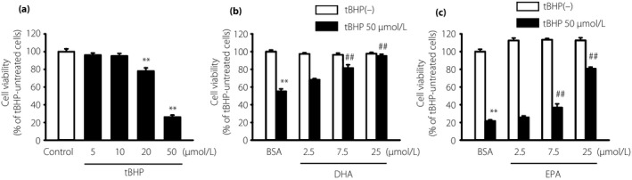
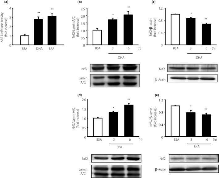
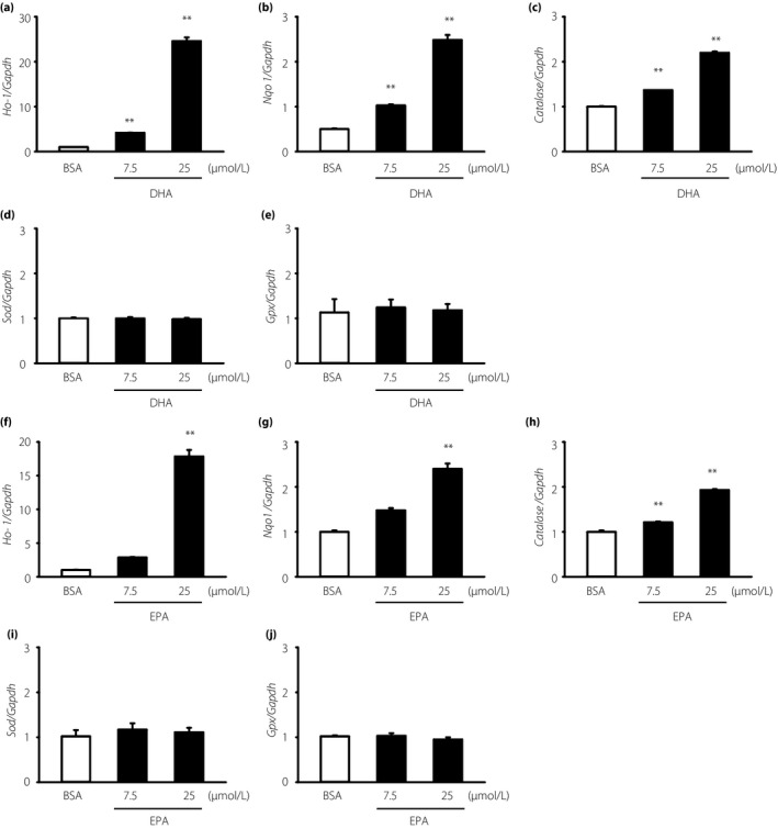
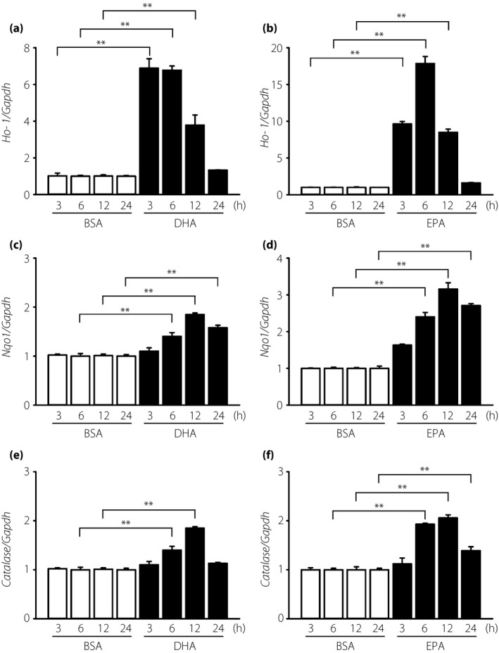
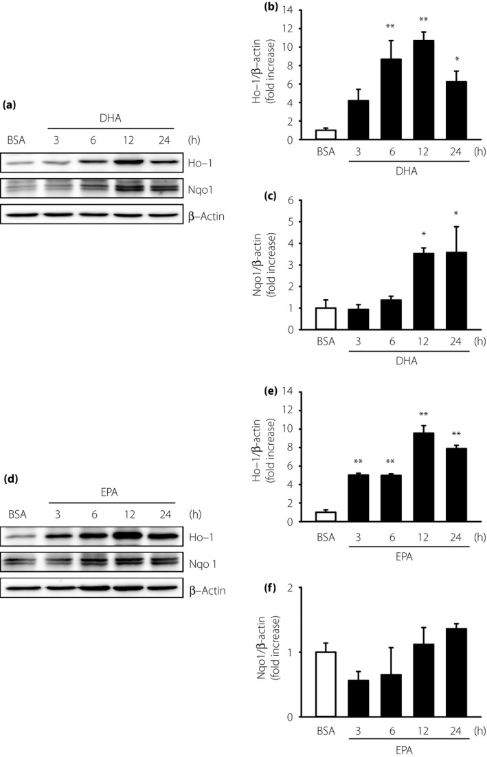
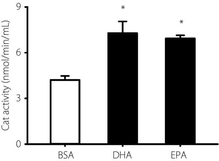
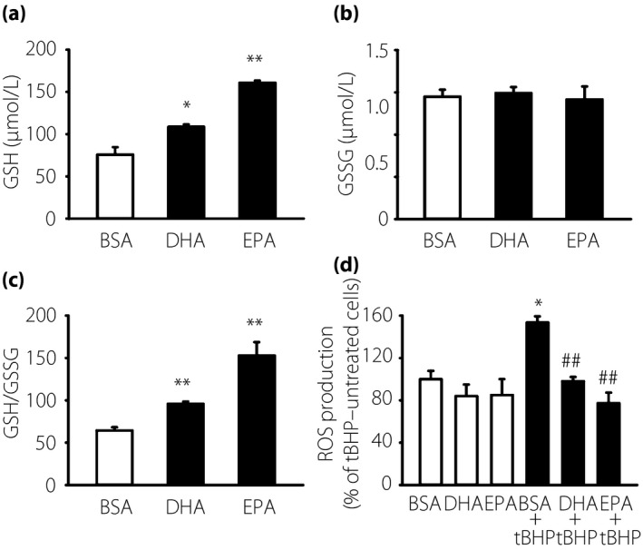
Treatment with tert-butyl hydroperoxide decreased cell viability dose-dependently. DHA or EPA pretreatment significantly alleviated tert-butyl hydroperoxide-induced cytotoxicity. DHA or EPA increased the messenger ribonucleic acid levels of Ho-1, nicotinamide adenine dinucleotide (phosphate) H quinone oxidoreductase 1 and catalase dose-dependently. Ho-1 protein level, catalase activity, Nrf2 promoter-luciferase activity and intracellular glutathione content were significantly increased by DHA and EPA.
These findings show that DHA and EPA can induce Ho-1 and catalase through Nrf2, thus protecting Schwann cells against oxidative stress. ω-3 PUFAs appear to exert their neuroprotective effect by increasing defense mechanisms against oxidative stress in diabetic neuropathies.
目的/引言:最近的研究表明,ω-3 多不饱和脂肪酸(ω-3 PUFAs)在血管中具有直接的抗氧化和抗炎作用;然而,ω-3 PUFAs 在许旺细胞中的作用仍未确定。
用 ω-3 PUFAs 二十二碳六烯酸(DHA)和二十碳五烯酸(EPA)孵育永生化的小鼠许旺(IMS32)细胞。使用实时逆转录聚合酶链反应鉴定几种抗氧化酶(血红素加氧酶-1[HO-1]、烟酰胺腺嘌呤二核苷酸[磷酸] H 醌氧化还原酶 1、过氧化氢酶、超氧化物歧化酶和谷胱甘肽过氧化物酶)的信使核糖核酸水平。使用 Western 印迹法评估 HO-1 和烟酰胺腺嘌呤二核苷酸[磷酸] H 醌氧化还原酶 1 蛋白水平。还使用 Western 印迹法对核部分的核因子(红细胞衍生 2)相关因子 2(Nrf2)进行定量。通过比色测定试剂盒测定过氧化氢酶活性和谷胱甘肽含量。通过双荧光素酶测定系统评估 Nrf2 启动子-荧光素酶活性。
叔丁基过氧化物处理剂量依赖性地降低细胞活力。DHA 或 EPA 预处理显著减轻叔丁基过氧化物诱导的细胞毒性。DHA 或 EPA 剂量依赖性地增加 HO-1、烟酰胺腺嘌呤二核苷酸[磷酸] H 醌氧化还原酶 1 和过氧化氢酶的信使核糖核酸水平。DHA 和 EPA 显著增加 HO-1 蛋白水平、过氧化氢酶活性、Nrf2 启动子-荧光素酶活性和细胞内谷胱甘肽含量。
这些发现表明,DHA 和 EPA 可以通过 Nrf2 诱导 HO-1 和过氧化氢酶,从而保护许旺细胞免受氧化应激。ω-3 PUFAs 似乎通过增加糖尿病神经病变中抗氧化应激的防御机制来发挥其神经保护作用。