Behzadi Ashkan Heshmatzadeh, Raza Syed Imran, Carrino John A, Kosmas Christos, Gholamrezanezhad Ali, Basques Kyle, Matcuk George R, Patel Jay, Jadvar Hossein
Department of Radiology, Weill Cornell Medical Center, 525 East 68th Street, New York, NY 10065, USA.
Department of Radiology, Weill Cornell Medical Center, 525 East 68th Street, New York, NY 10065, USA.
PET Clin. 2018 Oct;13(4):623-634. doi: 10.1016/j.cpet.2018.05.012. Epub 2018 Aug 17.
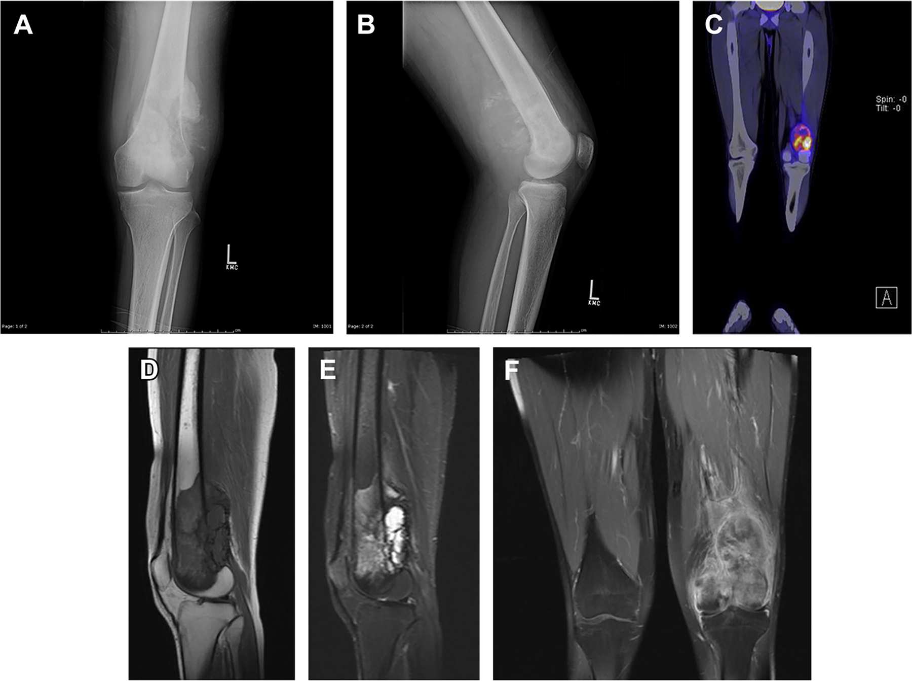
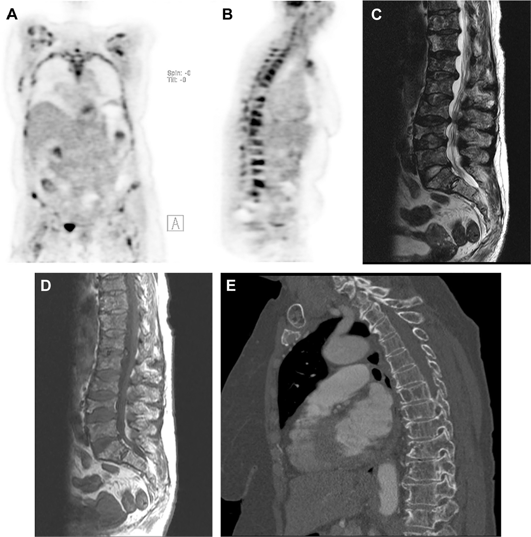
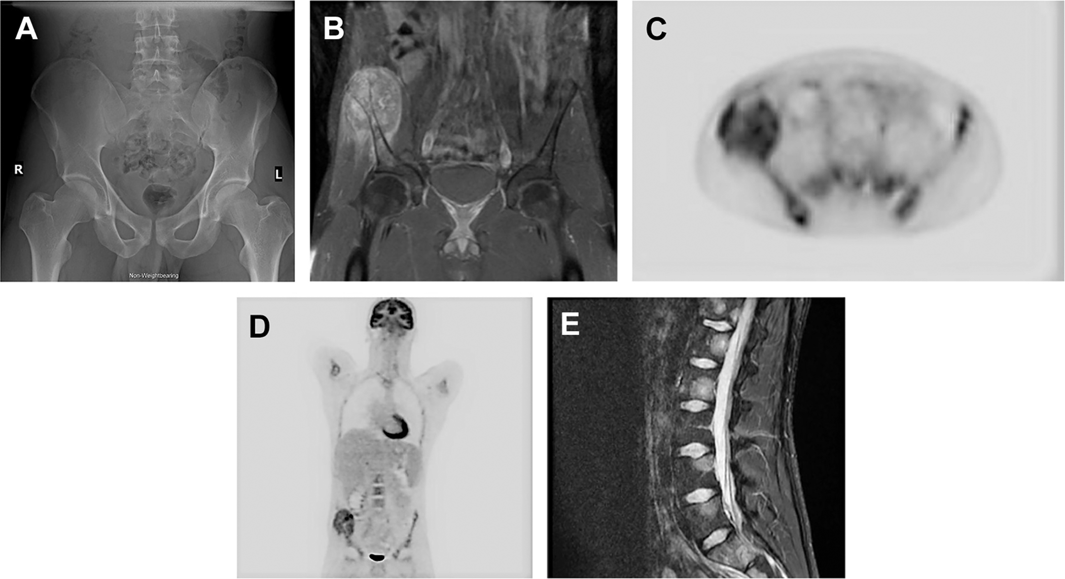
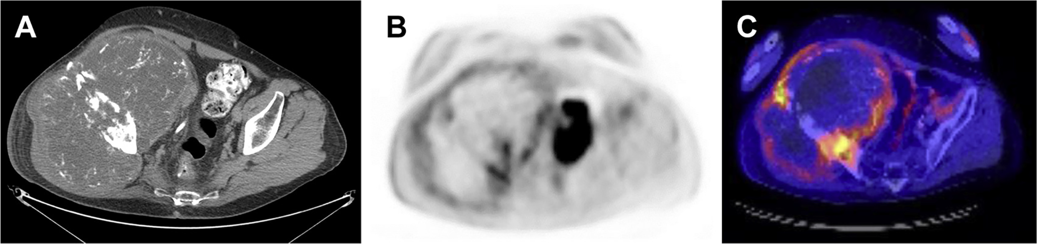
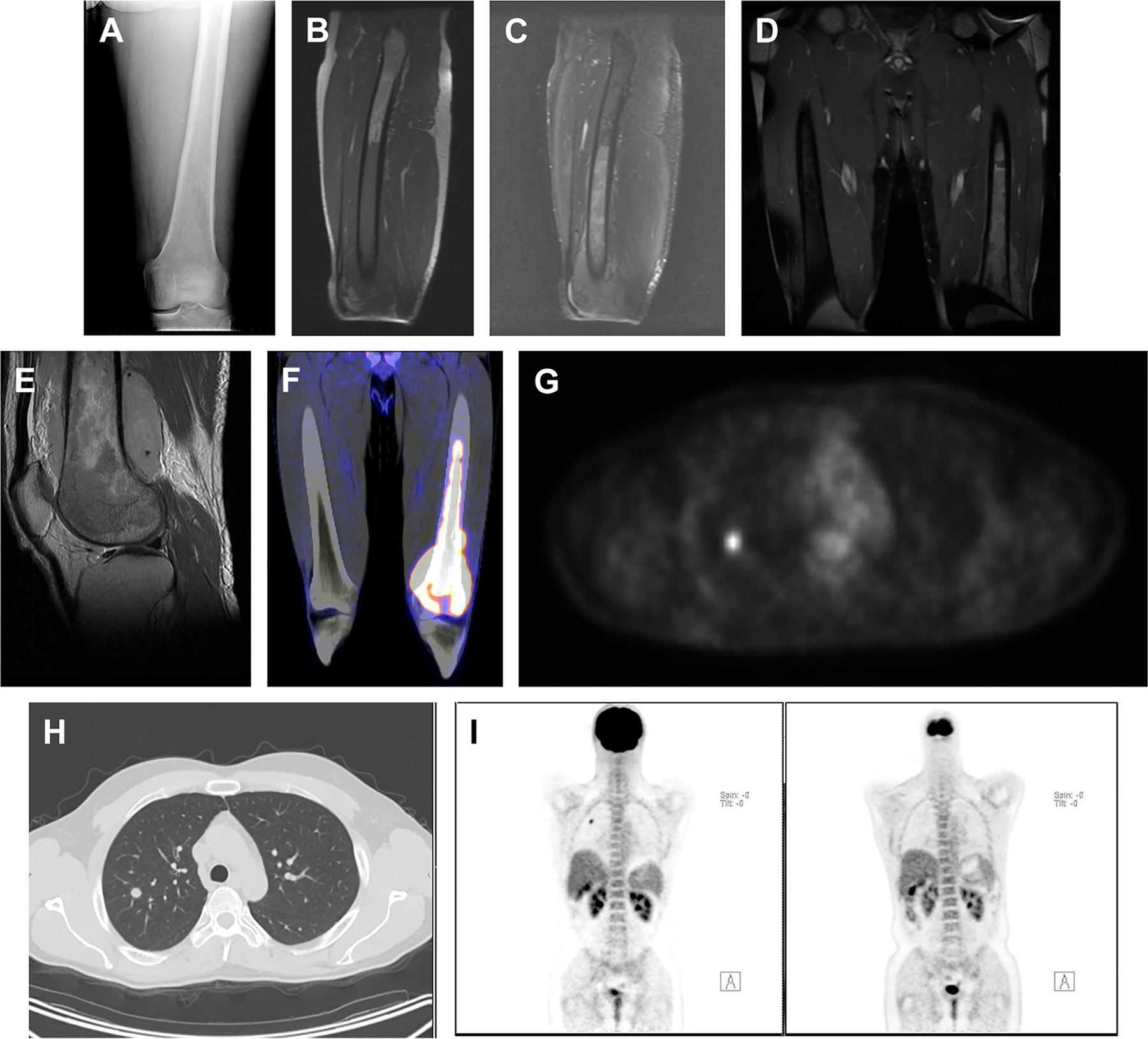
Primary bone malignancies are characterized with anatomic imaging. However, in recent years, there has been an increased interest in PET/computed tomography scanning and PET/MRI with fludeoxyglucose F 18 for evaluating and staging musculoskeletal neoplasms. These hybrid imaging modalities have shown promise largely owing to their high sensitivity, ability to perform more thorough staging, and ability to monitor treatment response. This article reviews the current role of PET/computed tomography scanning and PET/MRI in primary malignancies of bone, with an emphasis on imaging characteristics, clinical usefulness, and current limitations.
原发性骨恶性肿瘤通过解剖成像进行特征描述。然而,近年来,对于使用氟脱氧葡萄糖F 18进行正电子发射断层扫描/计算机断层扫描(PET/CT)以及正电子发射断层扫描/磁共振成像(PET/MRI)来评估和分期肌肉骨骼肿瘤的兴趣日益增加。这些混合成像模式已显示出前景,主要是由于其高灵敏度、能够进行更全面的分期以及能够监测治疗反应。本文综述了PET/CT扫描和PET/MRI在原发性骨恶性肿瘤中的当前作用,重点关注成像特征、临床实用性和当前局限性。