Department of Neurobiology and Anatomy, McGovern Medical School, University of Texas Health Science Center at Houston, Houston, Texas, United States of America.
The University of Texas MD Anderson Cancer Center UTHealth Graduate School of Biomedical Sciences, The University of Texas Health Science Center at Houston, Houston, Texas, United States America.
PLoS Biol. 2018 Sep 17;16(9):e2006169. doi: 10.1371/journal.pbio.2006169. eCollection 2018 Sep.
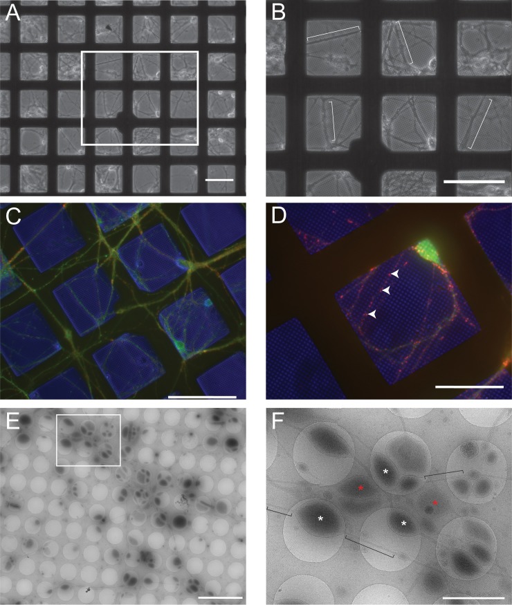
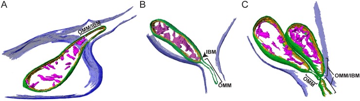
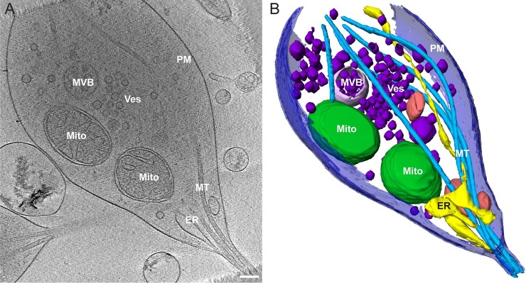
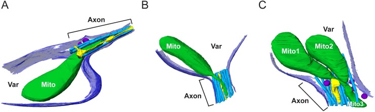
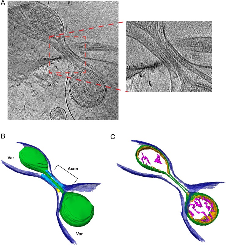
Neurons project axons to local and distal sites and can display heterogeneous morphologies with limited physical dimensions that may influence the structure of large organelles such as mitochondria. Using cryo-electron tomography (cryo-ET), we characterized native environments within axons and presynaptic varicosities to examine whether spatial restrictions within these compartments influence the morphology of mitochondria. Segmented tomographic reconstructions revealed distinctive morphological characteristics of mitochondria residing at the narrowed boundary between presynaptic varicosities and axons with limited physical dimensions (approximately 80 nm), compared to mitochondria in nonspatially restricted environments. Furthermore, segmentation of the tomograms revealed discrete organizations between the inner and outer membranes, suggesting possible independent remodeling of each membrane in mitochondria at spatially restricted axonal/varicosity boundaries. Thus, cryo-ET of mitochondria within axonal subcompartments reveals that spatial restrictions do not obstruct mitochondria from residing within them, but limited available space can influence their gross morphology and the organization of the inner and outer membranes. These findings offer new perspectives on the influence of physical and spatial characteristics of cellular environments on mitochondrial morphology and highlight the potential for remarkable structural plasticity of mitochondria to adapt to spatial restrictions within axons.
神经元投射轴突到局部和远端部位,并可显示出具有有限物理尺寸的异质形态,这可能影响到线粒体等大型细胞器的结构。使用冷冻电子断层扫描(cryo-ET),我们对轴突和突触前小泡内的天然环境进行了特征描述,以研究这些隔室内的空间限制是否会影响线粒体的形态。分段断层重建揭示了位于突触前小泡和轴突之间狭窄边界处的线粒体的独特形态特征,与非空间限制环境中的线粒体相比,这些线粒体具有有限的物理尺寸(约 80nm)。此外,断层图像的分割揭示了内外膜之间的离散组织,表明在空间受限的轴突/突触前小泡边界处,每个膜可能独立进行重塑。因此,对轴突亚区室中线粒体的 cryo-ET 显示,空间限制不会阻止线粒体存在于其中,但有限的可用空间会影响它们的总体形态和内外膜的组织。这些发现为细胞环境的物理和空间特征对线粒体形态的影响提供了新的视角,并强调了线粒体适应轴突内空间限制的显著结构可塑性的潜力。