Department of Integrative Structural and Computational Biology, The Scripps Research Institute , La Jolla, CA, USA.
Department of Molecular Medicine, The Scripps Research Institute , La Jolla, CA, USA.
J Cell Biol. 2023 Apr 3;222(4). doi: 10.1083/jcb.202204093. Epub 2023 Feb 14.
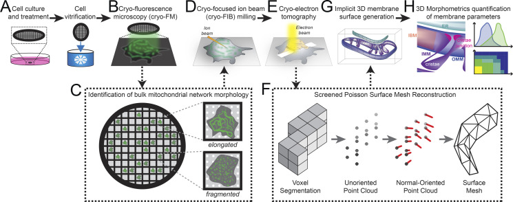
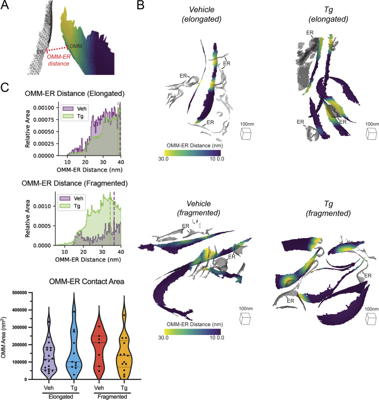
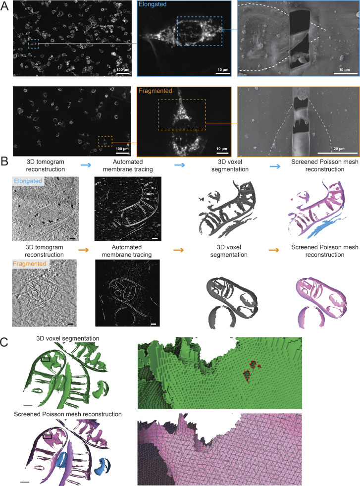
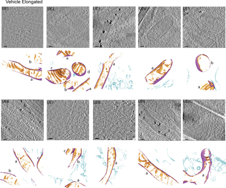
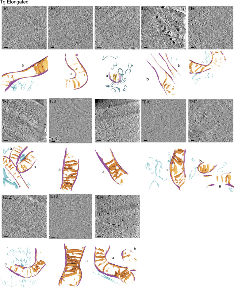
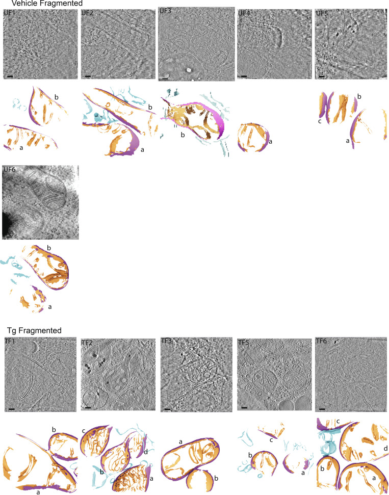
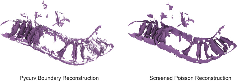
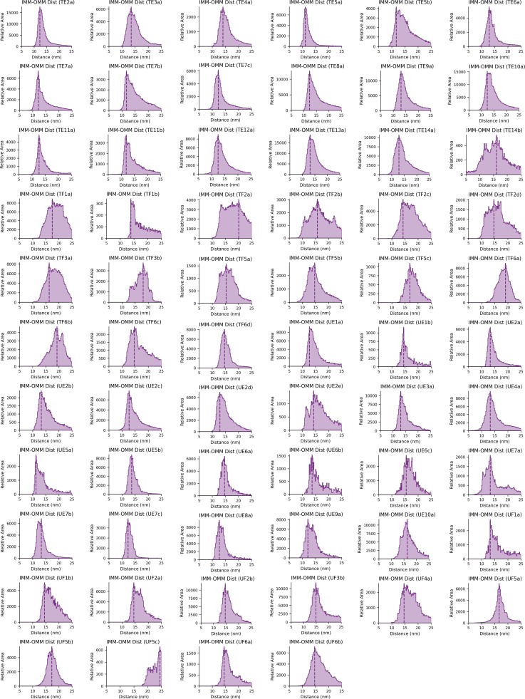
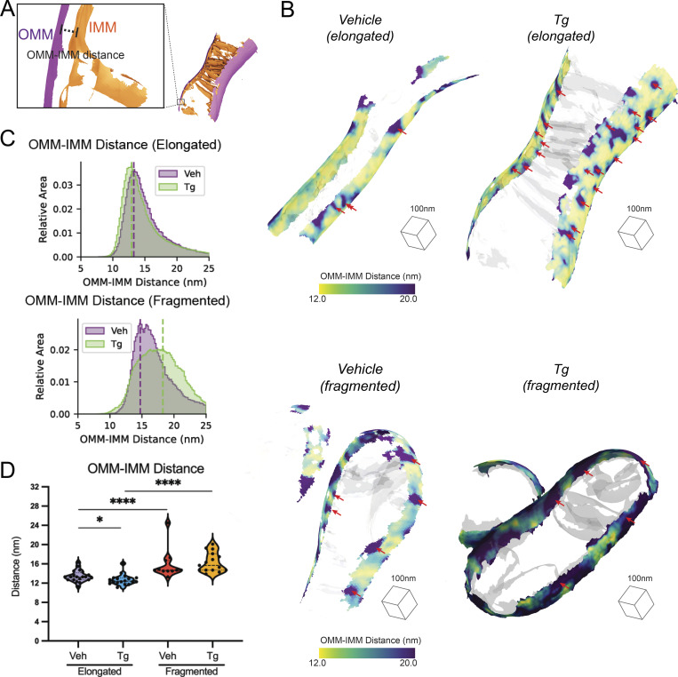
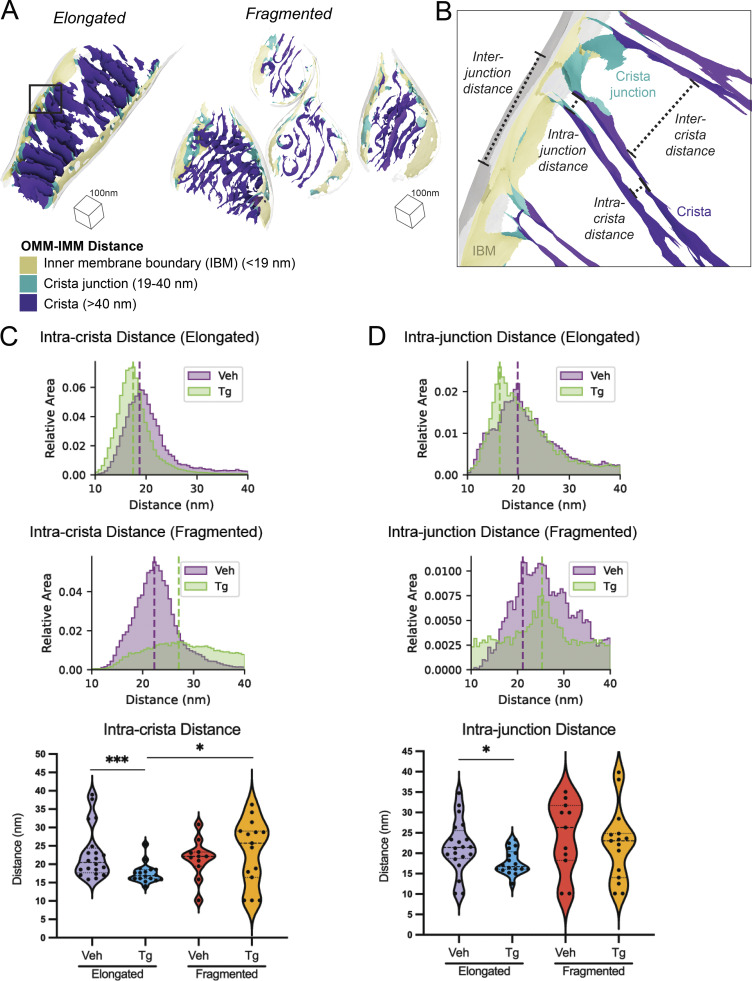
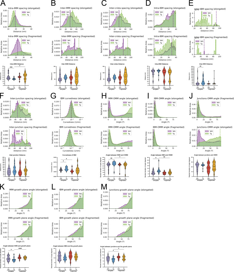
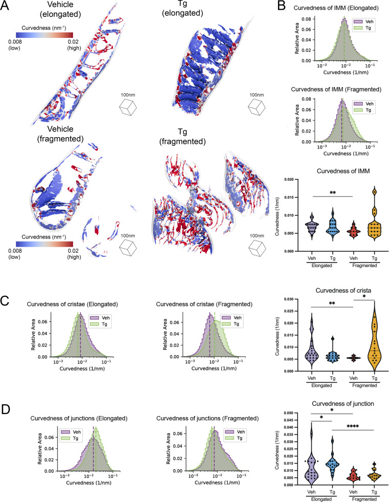
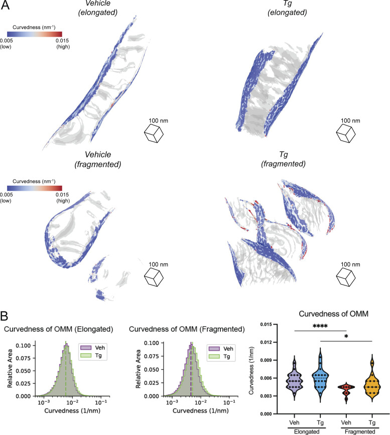
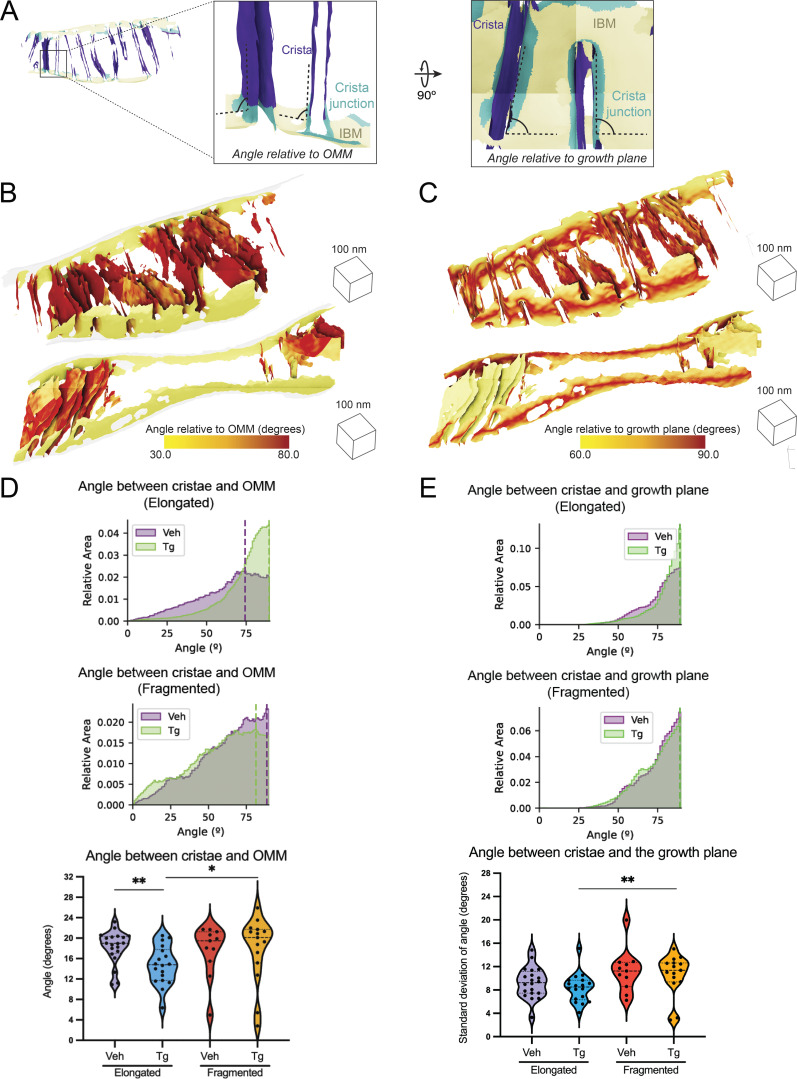
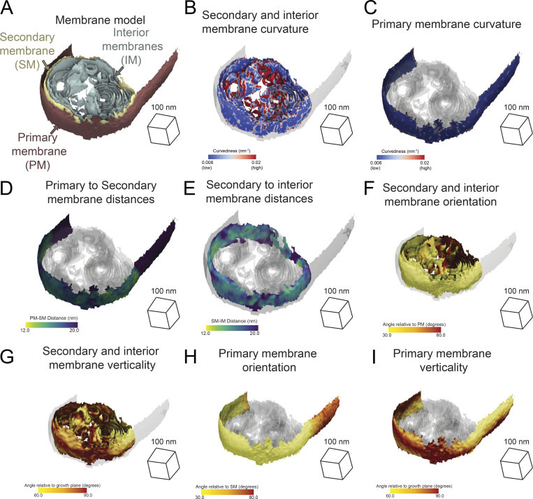
Cellular cryo-electron tomography (cryo-ET) enables three-dimensional reconstructions of organelles in their native cellular environment at subnanometer resolution. However, quantifying ultrastructural features of pleomorphic organelles in three dimensions is challenging, as is defining the significance of observed changes induced by specific cellular perturbations. To address this challenge, we established a semiautomated workflow to segment organellar membranes and reconstruct their underlying surface geometry in cryo-ET. To complement this workflow, we developed an open-source suite of ultrastructural quantifications, integrated into a single pipeline called the surface morphometrics pipeline. This pipeline enables rapid modeling of complex membrane structures and allows detailed mapping of inter- and intramembrane spacing, curvedness, and orientation onto reconstructed membrane meshes, highlighting subtle organellar features that are challenging to detect in three dimensions and allowing for statistical comparison across many organelles. To demonstrate the advantages of this approach, we combine cryo-ET with cryo-fluorescence microscopy to correlate bulk mitochondrial network morphology (i.e., elongated versus fragmented) with membrane ultrastructure of individual mitochondria in the presence and absence of endoplasmic reticulum (ER) stress. Using our pipeline, we demonstrate ER stress promotes adaptive remodeling of ultrastructural features of mitochondria including spacing between the inner and outer membranes, local curvedness of the inner membrane, and spacing between mitochondrial cristae. We show that differences in membrane ultrastructure correlate to mitochondrial network morphologies, suggesting that these two remodeling events are coupled. Our pipeline offers opportunities for quantifying changes in membrane ultrastructure on a single-cell level using cryo-ET, opening new opportunities to define changes in ultrastructural features induced by diverse types of cellular perturbations.
细胞冷冻电子断层扫描(cryo-ET)能够以亚纳米分辨率在其天然细胞环境中对细胞器进行三维重建。然而,对具有多形性的细胞器进行三维定量分析仍然具有挑战性,确定特定细胞扰动引起的观察到的变化的意义也具有挑战性。为了解决这个挑战,我们建立了一种半自动工作流程,用于分割细胞器膜并重建其底层表面几何形状的 cryo-ET。为了补充这个工作流程,我们开发了一套开源的超微结构定量分析套件,集成到一个名为表面形态计量学管道的单一管道中。该管道能够快速对复杂的膜结构进行建模,并允许将膜内和膜间的间隔、曲率和方向等详细信息映射到重建的膜网格上,突出显示在三维空间中难以检测到的微妙细胞器特征,并允许对许多细胞器进行统计比较。为了展示这种方法的优势,我们将 cryo-ET 与 cryo-fluorescence 显微镜相结合,以关联整体线粒体网络形态(即伸长与碎片化)与内质网(ER)应激存在和不存在时单个线粒体的膜超微结构。使用我们的管道,我们证明 ER 应激促进了线粒体超微结构特征的适应性重塑,包括内外膜之间的间隔、内膜的局部曲率和线粒体嵴之间的间隔。我们表明,膜超微结构的差异与线粒体网络形态相关,表明这两个重塑事件是耦合的。我们的管道提供了使用 cryo-ET 在单细胞水平上定量分析膜超微结构变化的机会,为定义不同类型的细胞扰动引起的超微结构特征变化开辟了新的机会。