Center for Ultrasound Molecular Imaging and Therapeutics, Department of Medicine and Heart and Vascular Institute, University of Pittsburgh School of Medicine and University of Pittsburgh Medical Center (UPMC), Pittsburgh, PA, 15261, USA.
Department of Bioengineering, School of Engineering, University of Pittsburgh, Pittsburgh, PA, 15261, USA.
Sci Rep. 2018 Sep 17;8(1):13918. doi: 10.1038/s41598-018-32235-2.
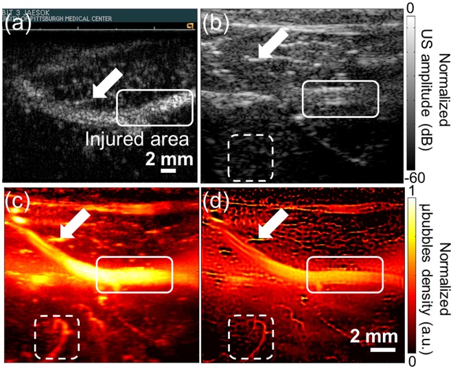
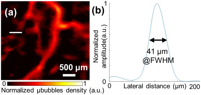
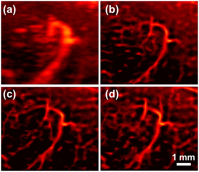
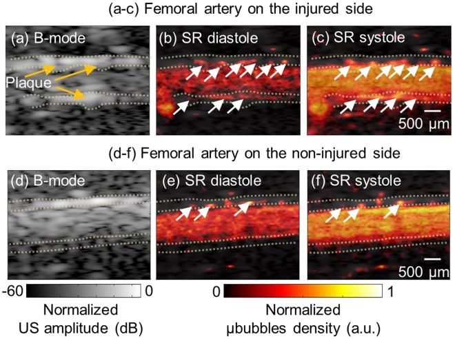
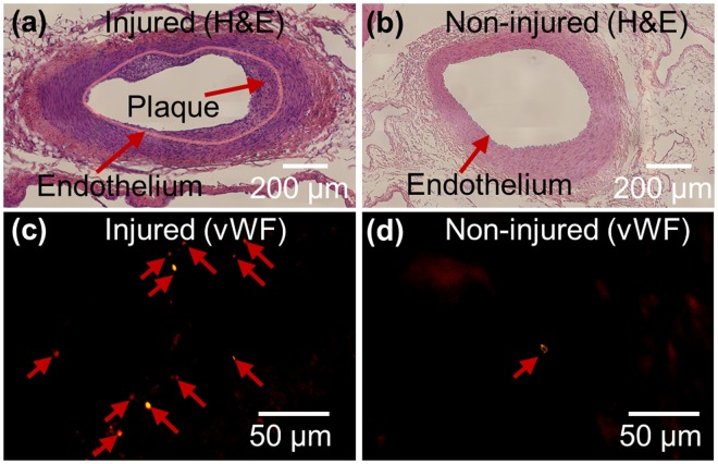
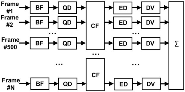
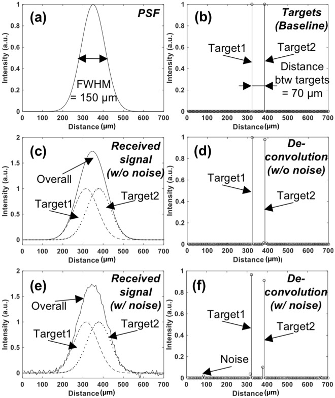
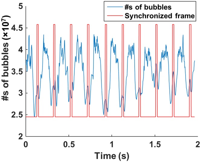
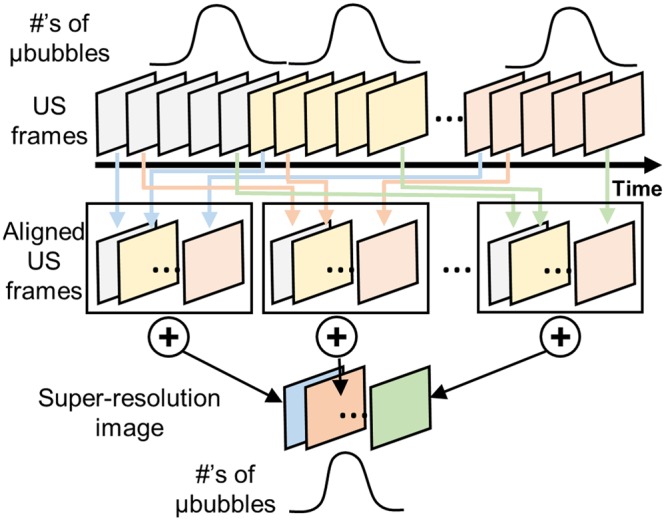
Traditional ultrasound imaging techniques are limited in spatial resolution to visualize angiogenic vasa vasorum that is considered as an important marker for atherosclerotic plaque progression and vulnerability. The recently introduced super-resolution imaging technique based on microbubble center localization has shown potential to achieve unprecedented high spatial resolution beyond the acoustic diffraction limit. However, a major drawback of the current super-resolution imaging approach is low temporal resolution because it requires a large number of imaging frames. In this study, a new imaging sequence and signal processing approach for super-resolution ultrasound imaging are presented to improve temporal resolution by employing deconvolution and spatio-temporal-interframe-correlation based data acquisition. In vivo feasibility of the developed technology is demonstrated and evaluated in imaging vasa vasorum in the rabbit atherosclerosis model. The proposed method not only identifies a tiny vessel with a diameter of 41 μm, 5 times higher spatial resolution than the acoustic diffraction limit at 7.7 MHz, but also significantly improves temporal resolution that allows for imaging vessels over cardiac motion.
传统的超声成像技术在空间分辨率上受到限制,无法可视化血管生成血管系统,而血管生成血管系统被认为是粥样斑块进展和易损性的一个重要标志物。最近引入的基于微泡中心定位的超分辨率成像技术具有超越声衍射极限实现前所未有的超高空间分辨率的潜力。然而,当前超分辨率成像方法的一个主要缺点是时间分辨率低,因为它需要大量的成像帧。在这项研究中,提出了一种新的成像序列和信号处理方法用于超分辨率超声成像,通过使用反卷积和基于时空帧间相关的数据采集来提高时间分辨率。在兔动脉粥样硬化模型中对所开发技术的体内可行性进行了演示和评估。所提出的方法不仅可以识别直径为 41μm 的微小血管,其空间分辨率比 7.7MHz 处的声衍射极限高 5 倍,而且还显著提高了时间分辨率,从而可以在心脏运动过程中对血管进行成像。