Botha S, Baitan D, Jungnickel K E J, Oberthür D, Schmidt C, Stern S, Wiedorn M O, Perbandt M, Chapman H N, Betzel C
Institute of Biochemistry and Molecular Biology, Chemistry Department, University of Hamburg, Martin-Luther-King Platz 6, 20146 Hamburg, Germany.
Laboratory for Structural Biology of Infection and Inflammation, c/o DESY, Building 22a, Notkestrasse 85, 22607 Hamburg, Germany.
IUCrJ. 2018 Aug 8;5(Pt 5):524-530. doi: 10.1107/S2052252518009223. eCollection 2018 Sep 1.
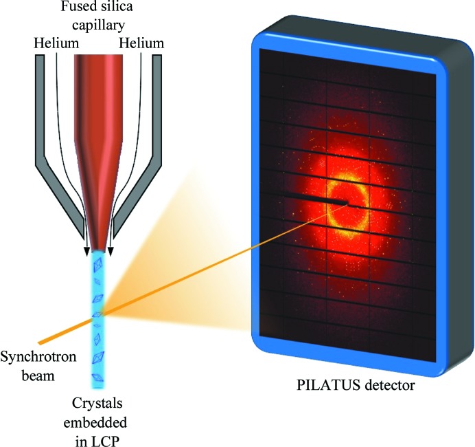
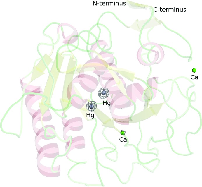
During the past few years, serial crystallography methods have undergone continuous development and serial data collection has become well established at high-intensity synchrotron-radiation beamlines and XFEL radiation sources. However, the application of experimental phasing to serial crystallography data has remained a challenging task owing to the inherent inaccuracy of the diffraction data. Here, a particularly gentle method for incorporating heavy atoms into micrometre-sized crystals utilizing lipidic cubic phase (LCP) as a carrier medium is reported. Soaking in LCP prior to data collection offers a new, efficient and gentle approach for preparing heavy-atom-derivative crystals directly before diffraction data collection using serial crystallography methods. This approach supports effective phasing by utilizing a reasonably low number of diffraction patterns. Using synchrotron radiation and exploiting the anomalous scattering signal of mercury for single isomorphous replacement with anomalous scattering (SIRAS) phasing resulted in high-quality electron-density maps that were sufficient for building a complete structural model of proteinase K at 1.9 Å resolution using automatic model-building tools.
在过去几年中,串行晶体学方法不断发展,串行数据采集在高强度同步辐射光束线和X射线自由电子激光(XFEL)辐射源处已得到广泛应用。然而,由于衍射数据固有的不准确性,将实验相位法应用于串行晶体学数据仍然是一项具有挑战性的任务。在此,报道了一种特别温和的方法,该方法利用脂质立方相(LCP)作为载体介质,将重原子引入微米级晶体中。在数据采集前浸泡在LCP中,为使用串行晶体学方法在衍射数据采集前直接制备重原子衍生晶体提供了一种新的、高效且温和的方法。这种方法通过使用数量合理的衍射图样来支持有效的相位确定。利用同步辐射并利用汞的反常散射信号进行单对映体置换反常散射(SIRAS)相位法,得到了高质量的电子密度图,这些图足以使用自动模型构建工具构建分辨率为1.9 Å的蛋白酶K完整结构模型。