Graduate Institute of Photonics and Optoelectronics, National Taiwan University, Taipei, Taiwan.
Department of Ophthalmology, National Taiwan University Hospital, Taipei, Taiwan.
Sci Rep. 2018 Sep 25;8(1):14349. doi: 10.1038/s41598-018-32814-3.
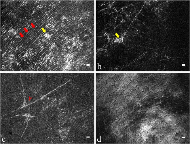
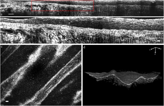
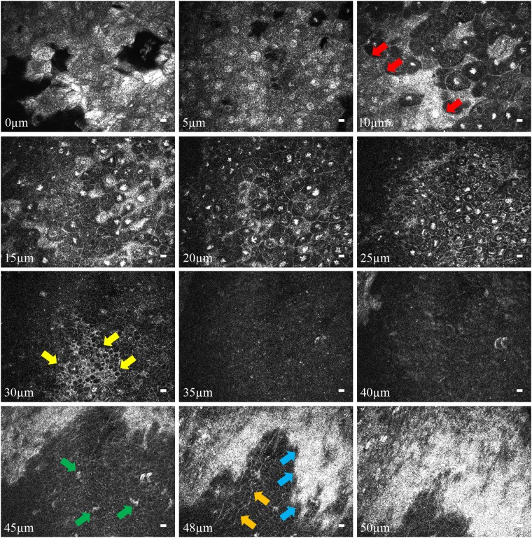
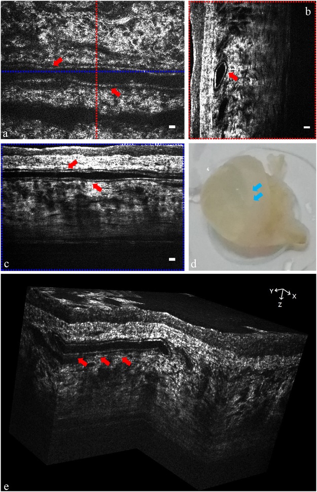
Accurate diagnosis of corneal pathology and morphological identification of different corneal layers require clear delineation of corneal three-dimensional structures and en face or cross-sectional imaging of palisade of Vogt (POV), neovascularization (NV) or corneal nerves. Here we report a prototype of full-field optical coherence tomography (FF-OCT) system with isotropic sub-micron spatial resolution in the en face and cross-sectional views. It can also provide three-dimensional reconstructed images and a large field of view (FOV) by stitching tomograms side by side. We validated the imaging power of this prototype in in vivo rat and rabbit eyes, and quantified anatomical characteristics such as corneal layer thickness, endothelial cell density and the intensity profile of different layers. This FF-OCT delineated the ridge-like structure of POV, corneal nerve bundles, and conjunctival vessels in rat eyes. It also clearly identified the vessel walls and red blood cells in rabbit model of corneal NV. The findings provided by this FF-OCT are expected to facilitate corneal disease diagnosis and treatment.
准确诊断角膜病变和对不同角膜层的形态学识别需要清晰地描绘角膜的三维结构,并对视盘栅(POV)、新生血管(NV)或角膜神经的面内或横截面成像。在这里,我们报告了一种具有各向同性亚微米空间分辨率的全场光学相干断层扫描(FF-OCT)系统的原型,它可以通过并排拼接断层扫描来提供三维重建图像和大视场(FOV)。我们在体内大鼠和兔眼验证了该原型的成像能力,并量化了角膜层厚度、内皮细胞密度和不同层的强度分布等解剖特征。该 FF-OCT 描绘了大鼠眼中 POV 的脊状结构、角膜神经束和结膜血管。它还清楚地识别了兔角膜 NV 模型中的血管壁和红细胞。该 FF-OCT 提供的结果有望促进角膜疾病的诊断和治疗。