Department of Ophthalmology, National Taiwan University Hospital, Taipei, Taiwan.
Graduate Institute of Clinical Medicine, College of Medicine, National Taiwan University, Taipei, Taiwan.
Sci Rep. 2021 Feb 10;11(1):3492. doi: 10.1038/s41598-021-82178-4.
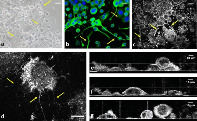
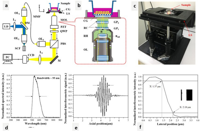
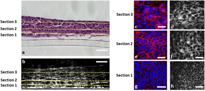
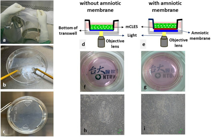
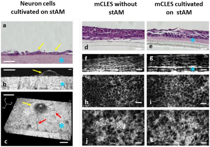
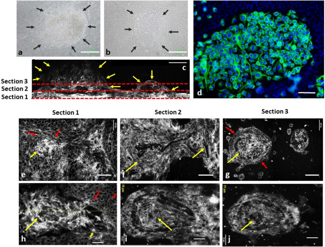
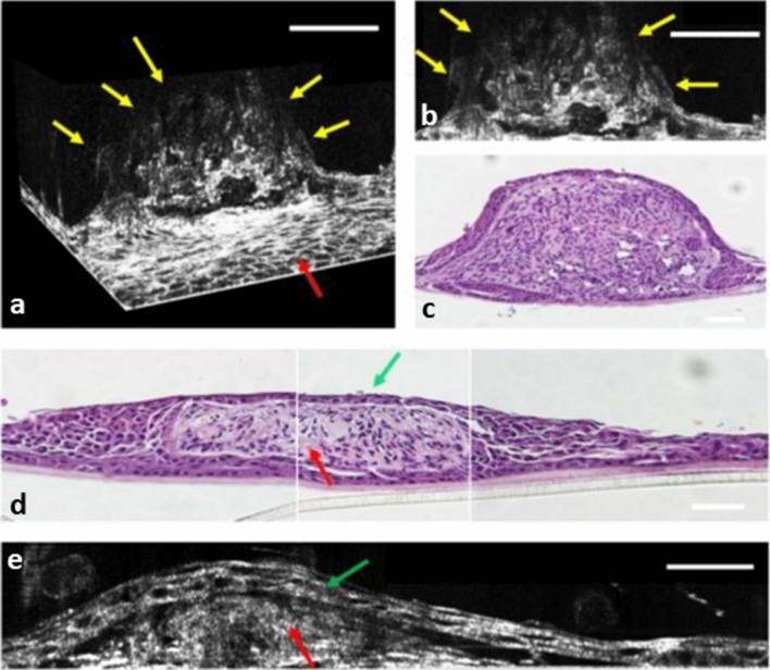
Three-dimensional (3D) configuration of in vitro cultivated cells has been recognised as a valuable tool in developing stem cell and cancer cell therapy. However, currently available imaging approaches for live cells have drawbacks, including unsatisfactory resolution, lack of cross-sectional and 3D images, and poor penetration of multi-layered cell products, especially when cells are cultivated on semitransparent carriers. Herein, we report a prototype of a full-field optical coherence tomography (FF-OCT) system with isotropic submicron spatial resolution in en face and cross-sectional views that provides a label-free, non-invasive platform with high-resolution 3D imaging. We validated the imaging power of this prototype by examining (1) cultivated neuron cells (N2A cell line); (2) multilayered, cultivated limbal epithelial sheets (mCLESs); (3) neuron cells (N2A cell line) and mCLESs cultivated on a semitransparent amniotic membrane (stAM); and (4) directly adherent colonies of neuron-like cells (DACNs) covered by limbal epithelial cell sheets. Our FF-OCT exhibited a penetrance of up to 150 μm in a multilayered cell sheet and displayed the morphological differences of neurons and epithelial cells in complex coculture systems. This FF-OCT is expected to facilitate the visualisation of cultivated cell products in vitro and has a high potential for cell therapy and translational medicine research.
三维(3D)培养细胞结构已被认为是开发干细胞和癌细胞治疗的有价值的工具。然而,目前用于活细胞的成像方法存在缺陷,包括分辨率不理想、缺乏横截面和 3D 图像以及多层细胞产品的穿透性差,尤其是当细胞在半透明载体上培养时。在此,我们报告了一种具有各向同性亚微米空间分辨率的全场光学相干断层扫描(FF-OCT)系统原型,该系统在共面和横截面视图中提供了无标记、非侵入性的高分辨率 3D 成像平台。我们通过检查(1)培养的神经元细胞(N2A 细胞系);(2)多层培养的角膜缘上皮片(mCLESs);(3)神经元细胞(N2A 细胞系)和培养在半透明羊膜(stAM)上的 mCLESs;以及(4)直接附着的神经元样细胞(DACNs)集落覆盖的角膜缘上皮细胞片,验证了该原型的成像能力。我们的 FF-OCT 在多层细胞片中的穿透深度可达 150μm,并显示了复杂共培养系统中神经元和上皮细胞的形态差异。这种 FF-OCT 有望促进体外培养细胞产物的可视化,并且在细胞治疗和转化医学研究中有很高的应用潜力。