Computer Aided Medical Procedures, Technical University of Munich, 85748, Garching, Germany.
Chair of Biomedical Physics, Department of Physics and Munich School of BioEngineering, Technical University of Munich, 85748, Garching, Germany.
Sci Rep. 2018 Sep 25;8(1):14345. doi: 10.1038/s41598-018-32023-y.
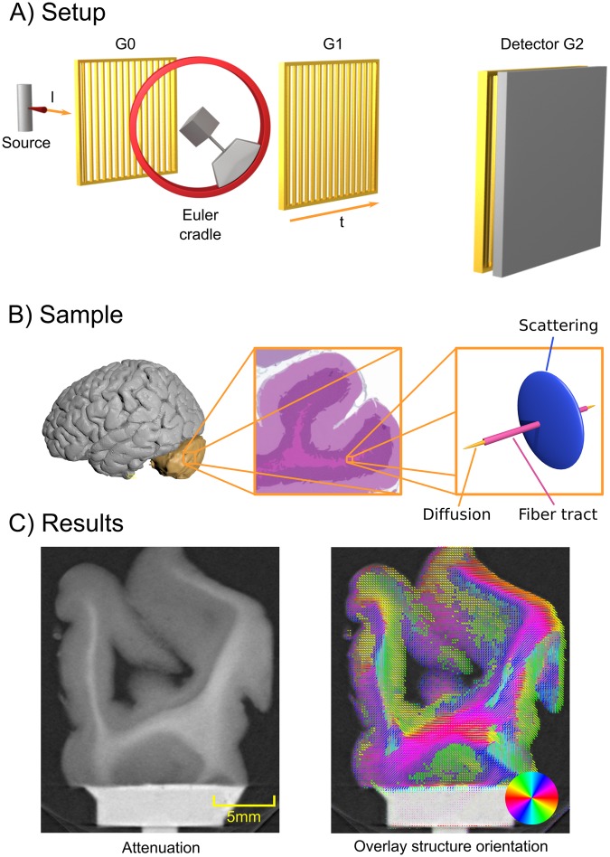
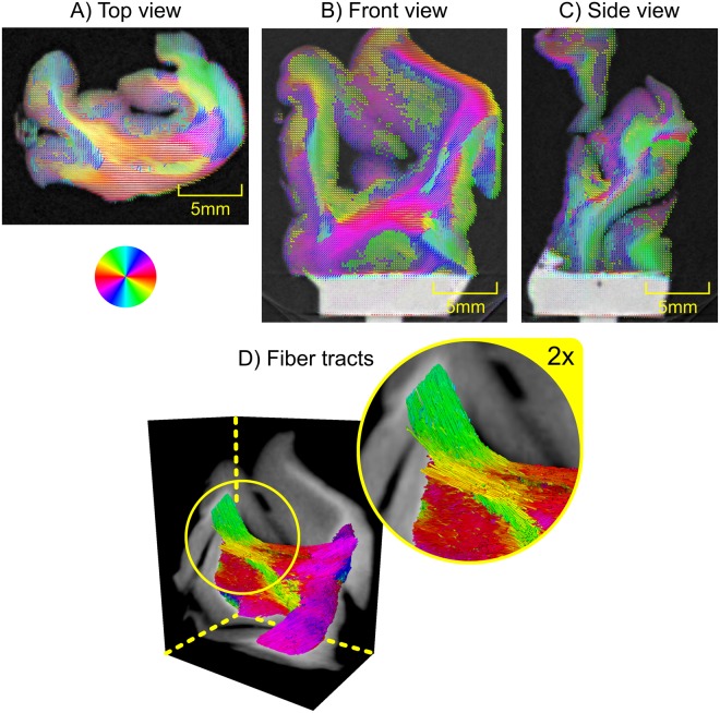
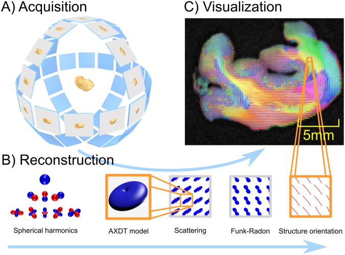
To understand the interaction of different parts of the human brain it is essential to know how they are connected. Such connections are predominantly related to the brain's white matter, which forms the neuronal pathways, the axons. These axons, also referred to as nerve fibers, have a size on the micrometer scale and are therefore too small to be imaged by standard X-ray systems. In this paper, we use a grating interferometer and a method based on Anisotropic X-ray Dark-field Tomography (AXDT) with the goal to generate a three-dimensional tomographic reconstruction of these functional structures. A first preclinical survey shows that we successfully reconstruct the orientations of the brain fibers connectivity with our approach.
为了理解人类大脑不同部分的相互作用,了解它们是如何连接的至关重要。这种连接主要与大脑的白质有关,白质形成了神经元通路,即轴突。这些轴突也被称为神经纤维,其大小在微米范围内,因此太小而无法用标准 X 射线系统成像。在本文中,我们使用了一个光栅干涉仪和一种基于各向异性 X 射线暗场层析成像(AXDT)的方法,目的是对这些功能结构进行三维层析重建。初步的临床前研究表明,我们成功地用我们的方法重建了大脑纤维连接的方向。