Fan Yang, Chen Zhilu, Pathak Janak L, Carneiro Ana M D, Chung Chang Y
School of Pharmaceutical Science and Technology, Tianjin University, Tianjin, China.
Department of Pharmacology, Vanderbilt University Medical Center, Vanderbilt University, Nashville, TN, United States.
Front Cell Neurosci. 2018 Sep 11;12:309. doi: 10.3389/fncel.2018.00309. eCollection 2018.
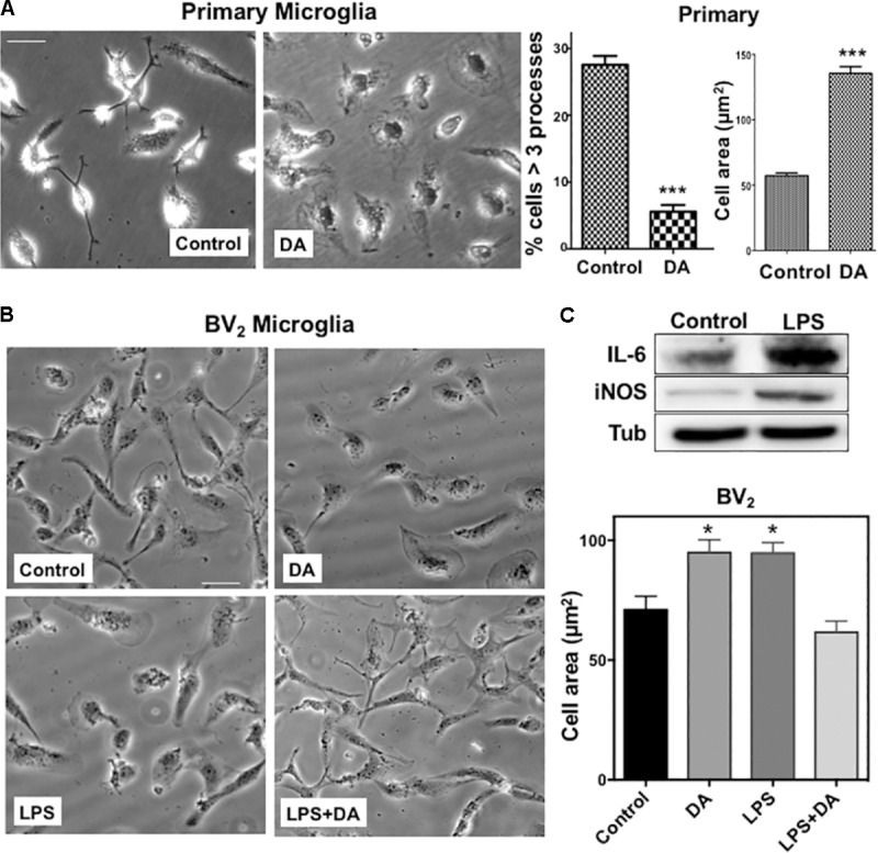
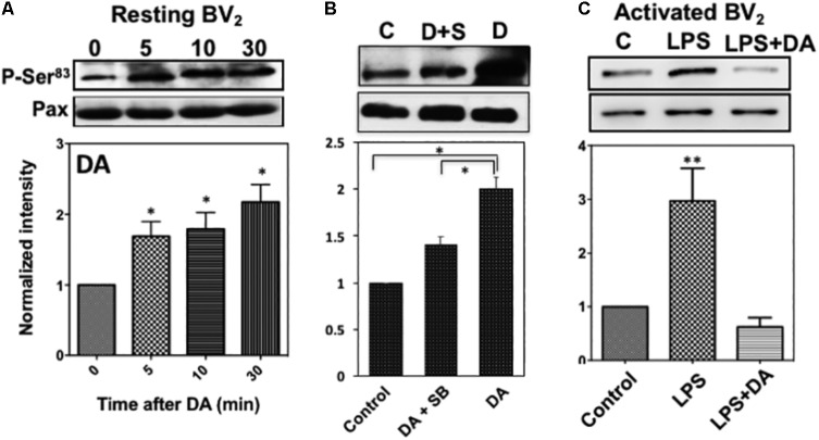
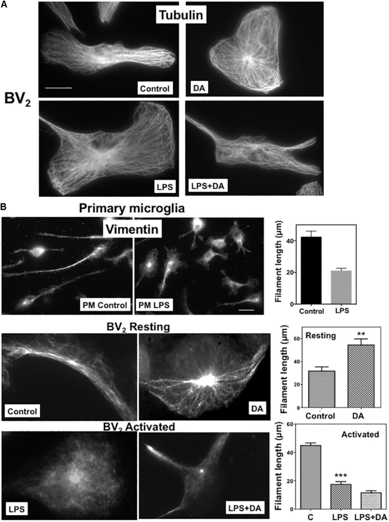
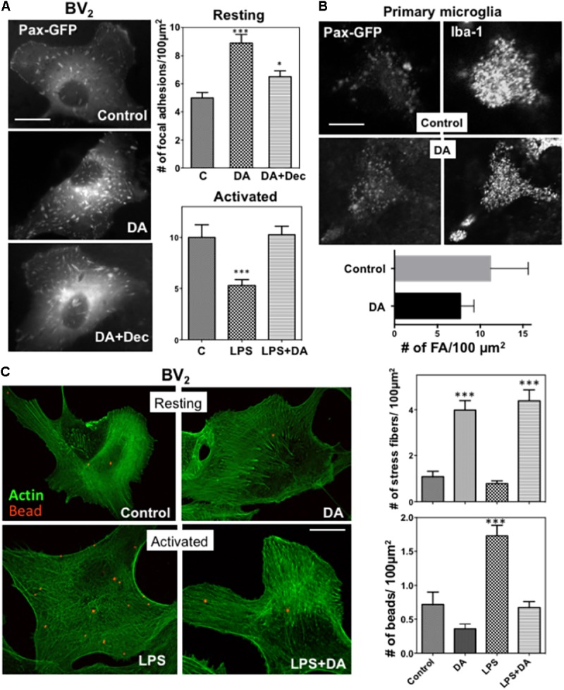
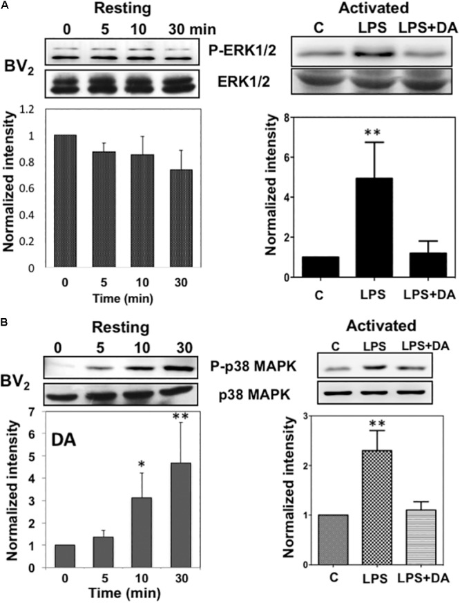
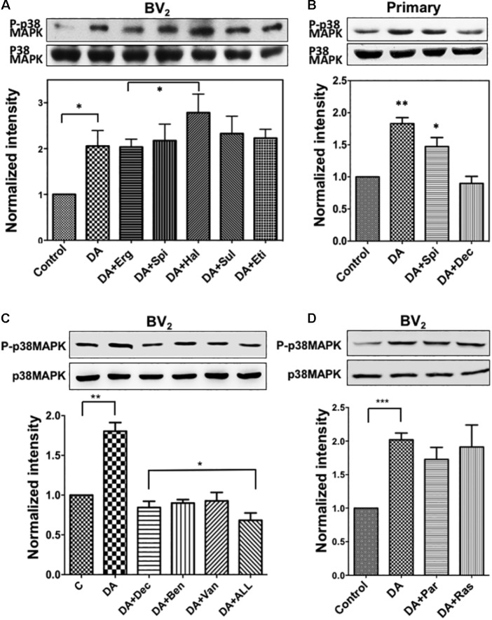
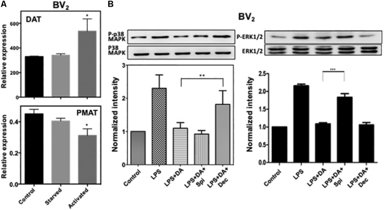
Microglia, the immune competent cells of the central nervous system (CNS), normally exist in a resting state characterized by a ramified morphology with many processes, and become activated to amoeboid morphology in response to brain injury, infection, and a variety of neuroinflammatory stimuli. Many studies focused on how neurotransmitters affect microglia activation in pathophysiological circumstances. In this study, we tried to gain mechanistic insights on how dopamine (DA) released from neurons modulates cellular functions of resting and activated microglia. DA induced the reduction of the number of cellular processes, the increase of cell adhesion/spreading, and the increase of vimentin filaments in resting primary and BV microglia. In contrast to resting cells, DA downregulated the cell spreading and phagocytosis of microglia activated by LPS. DA also significantly downregulated ERK1/2 phosphorylation in activated microglia, but not in resting microglia. Downregulation of ERK1/2 by DA in activated microglia required receptor signaling. In contrast, we found a significant increase of p38MAPK activity by DA treatment in resting, but not in activated microglia. These latter effects required the uptake of DA through the high-affinity transporter but did not require receptor signaling. Activation of p38MAPK resulted in the increase of focal adhesion number via phosphorylation of paxillin at Ser. These results indicate that DA might have a differential, depending upon the activation stage of microglia, impact on cellular functions such as adhesion and phagocytosis.
小胶质细胞是中枢神经系统(CNS)具有免疫活性的细胞,通常以具有许多分支的分支形态处于静息状态,并在脑损伤、感染和各种神经炎症刺激下被激活为阿米巴样形态。许多研究聚焦于神经递质在病理生理情况下如何影响小胶质细胞的激活。在本研究中,我们试图深入了解神经元释放的多巴胺(DA)如何调节静息和激活的小胶质细胞的细胞功能。DA诱导原代静息小胶质细胞和BV小胶质细胞的细胞分支数量减少、细胞黏附/铺展增加以及波形蛋白丝增加。与静息细胞相反,DA下调了由脂多糖激活的小胶质细胞的细胞铺展和吞噬作用。DA还显著下调激活的小胶质细胞中ERK1/2的磷酸化,但对静息小胶质细胞无此作用。DA在激活的小胶质细胞中对ERK1/2的下调需要受体信号传导。相反,我们发现DA处理使静息小胶质细胞中的p38MAPK活性显著增加,但对激活的小胶质细胞无此作用。后一种作用需要通过高亲和力转运体摄取DA,但不需要受体信号传导。p38MAPK的激活导致桩蛋白在Ser位点磷酸化,从而增加黏着斑数量。这些结果表明,DA可能根据小胶质细胞的激活阶段,对诸如黏附和吞噬等细胞功能产生不同影响。