Mukai Takeo, Di Martino Elena, Tsuji Shunichiro, Blomgren Klas, Nagamura-Inoue Tokiko, Ådén Ulrika
Department of Women's and Children's Health, Karolinska Institutet, Stockholm, Sweden.
Department of Cell Processing and Transfusion, The Institute of Medical Science, The University of Tokyo, Bunkyo City, Japan.
Cell Death Discov. 2021 Mar 15;7(1):46. doi: 10.1038/s41420-021-00436-w.
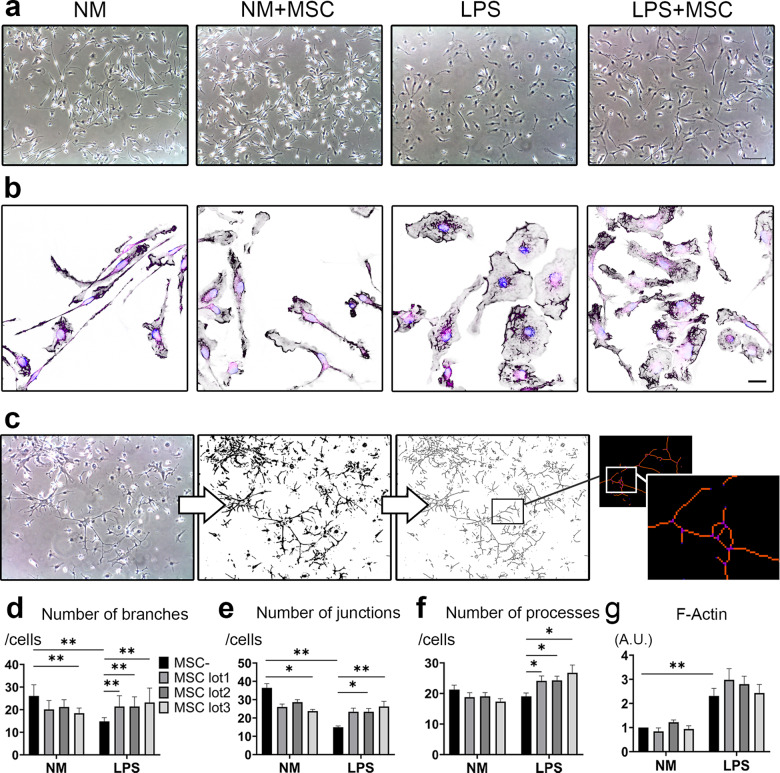
Microglia are the immune cells in the central nervous system surveying environment and reacting to various injuries. Activated microglia may cause impaired synaptic plasticity, therefore modulating and restoring them to neutral phenotype is crucial to counteract a pro-inflammatory, neurotoxic state. In this study, we focused on elucidating whether human umbilical cord (UC) -derived mesenchymal stromal cells (MSCs) can exert immunomodulatory effect and change the phenotype of activated microglia. Primary culture of microglia was activated by lipopolysaccharide (LPS) and was co-cultured with three lots of MSCs. We investigated immunomodulation, actin dynamics and phagocytic capacity of activated microglia, and examined change of Rho GTPase in microglia as the mechanism. MSCs suppressed the expression of IL-1β and pNFκB in LPS-activated microglia, and conversely elevated the expression of IL-1β in resting-surveying microglia with lot-to-lot variation. Morphological and phagocytotic analyses revealed that LPS stimulation significantly increased active Rho GTPase, Rac1, and Cdc42 levels in the microglia, and their morphology changed to amoeboid in which F-actin spread with ruffle formation. The F-actin spreading persisted after removal of LPS stimulation and reduced phagocytosis. On the other hand, MSC co-culture induced bimodal increase in active Rac1 and Cdc42 levels in LPS-activated microglia. Moreover, extended ruffles of F-actin shrinked and concentrated to form an actin ring, thereby restoring phagocytosis. We confirmed inhibition of the PI3K/Akt pathway attenuated F-actin dynamics and phagocytosis restored by MSCs. Overall, we demonstrated that MSCs immunomodulated microglia with lot-to-lot variation, and changed the phenotype of LPS-activated microglia restoring actin dynamics and phagocytosis by increase of active Rho GTPase.
小胶质细胞是中枢神经系统中的免疫细胞,负责监测环境并对各种损伤做出反应。活化的小胶质细胞可能导致突触可塑性受损,因此调节并使其恢复到中性表型对于对抗促炎、神经毒性状态至关重要。在本研究中,我们着重阐明人脐带(UC)来源的间充质基质细胞(MSC)是否能发挥免疫调节作用并改变活化小胶质细胞的表型。小胶质细胞原代培养物经脂多糖(LPS)活化后,与三批MSC共培养。我们研究了活化小胶质细胞的免疫调节、肌动蛋白动力学和吞噬能力,并将小胶质细胞中Rho GTP酶的变化作为机制进行研究。MSC抑制LPS活化的小胶质细胞中IL-1β和pNFκB的表达,相反,在静息监测的小胶质细胞中,不同批次的MSC会使IL-1β表达升高。形态学和吞噬分析显示,LPS刺激显著增加了小胶质细胞中活性Rho GTP酶、Rac1和Cdc42的水平,其形态转变为阿米巴样,F-肌动蛋白伸展并形成褶皱。去除LPS刺激后,F-肌动蛋白的伸展持续存在,吞噬作用降低。另一方面,MSC共培养导致LPS活化的小胶质细胞中活性Rac1和Cdc42水平出现双峰增加。此外,F-肌动蛋白的延长褶皱收缩并集中形成肌动蛋白环,从而恢复吞噬作用。我们证实PI3K/Akt通路的抑制减弱了F-肌动蛋白动力学,而MSC恢复了吞噬作用。总体而言,我们证明了MSC对小胶质细胞具有批次间差异的免疫调节作用,并通过增加活性Rho GTP酶改变了LPS活化小胶质细胞的表型,恢复了肌动蛋白动力学和吞噬作用。