• 文献检索
  • 文档翻译
  • 深度研究
  • 学术资讯
  • Suppr Zotero 插件Zotero 插件
  • 邀请有礼
  • 套餐&价格
  • 历史记录
应用&插件
Suppr Zotero 插件Zotero 插件浏览器插件Mac 客户端Windows 客户端微信小程序
定价
高级版会员购买积分包购买API积分包
服务
文献检索文档翻译深度研究API 文档MCP 服务
关于我们
关于 Suppr公司介绍联系我们用户协议隐私条款
关注我们

Suppr 超能文献

核心技术专利:CN118964589B侵权必究
粤ICP备2023148730 号-1Suppr @ 2026

文献检索

告别复杂PubMed语法,用中文像聊天一样搜索,搜遍4000万医学文献。AI智能推荐,让科研检索更轻松。

立即免费搜索

文件翻译

保留排版,准确专业,支持PDF/Word/PPT等文件格式,支持 12+语言互译。

免费翻译文档

深度研究

AI帮你快速写综述,25分钟生成高质量综述,智能提取关键信息,辅助科研写作。

立即免费体验

轮状病毒侵入细胞过程中钙离子流失的可视化。

Visualization of Calcium Ion Loss from Rotavirus during Cell Entry.

机构信息

Laboratory of Molecular Medicine, Boston Children's Hospital, Harvard Medical School, Boston, Massachusetts, USA.

Department of Chemistry, University of Chicago, Chicago, Illinois, USA.

出版信息

J Virol. 2018 Nov 27;92(24). doi: 10.1128/JVI.01327-18. Print 2018 Dec 15.

DOI:10.1128/JVI.01327-18
PMID:30258012
原文链接:https://pmc.ncbi.nlm.nih.gov/articles/PMC6258952/
Abstract

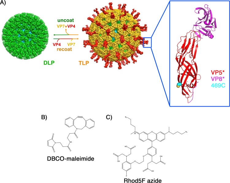
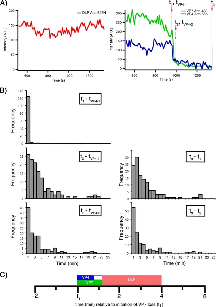
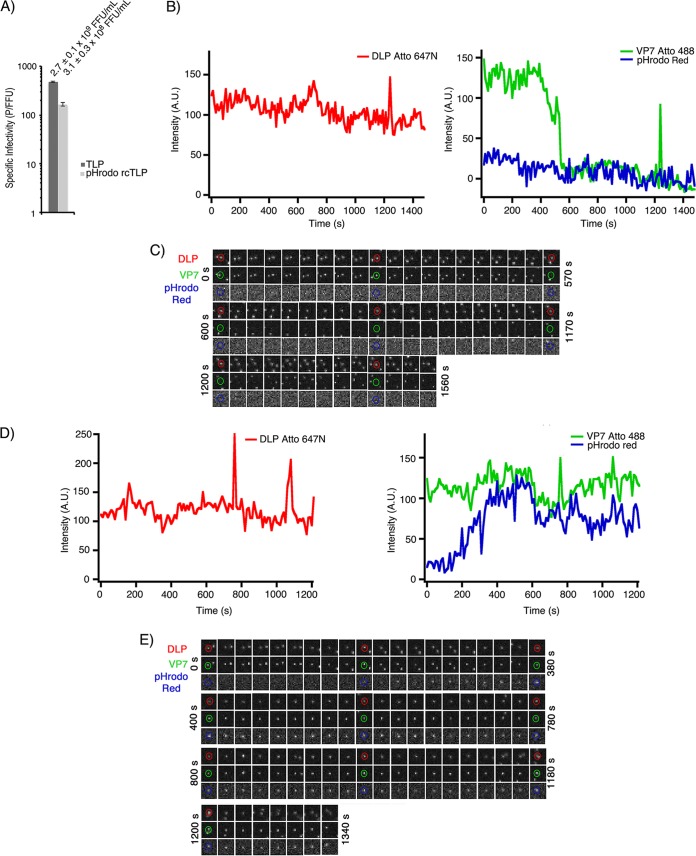
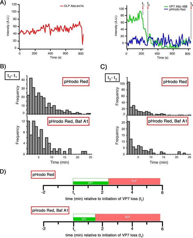
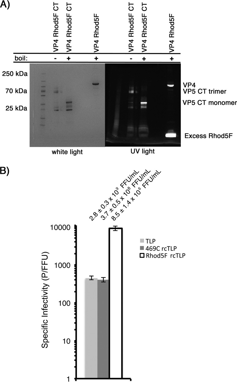
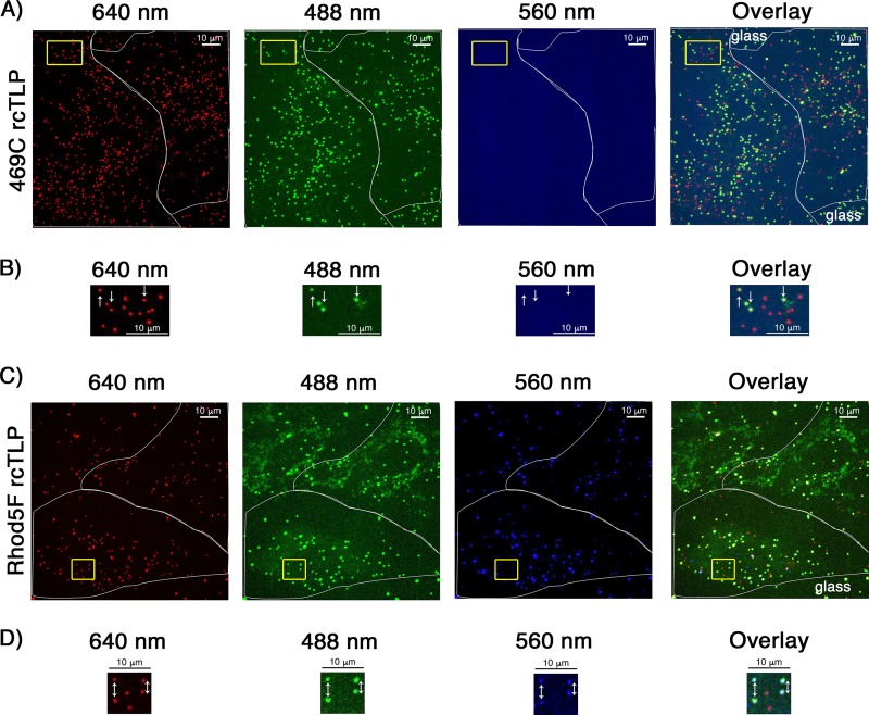
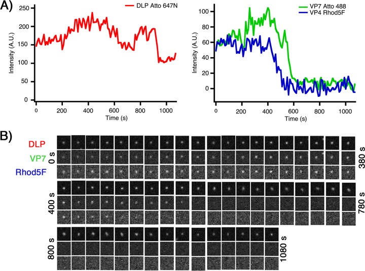
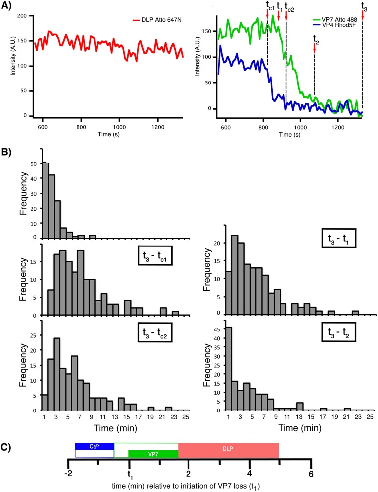
Bound calcium ions stabilize many nonenveloped virions. Loss of Ca from these particles appears to be a regulated part of entry or uncoating. The outer layer of an infectious rotavirus triple-layered particle (TLP) comprises a membrane-interacting protein (VP4) anchored by a Ca-stabilized protein (VP7). Membrane-coupled conformational changes in VP4 (cleaved to VP8* and VP5*) and dissociation of VP4 and VP7 accompany penetration of the double-layered inner capsid particle (DLP) into the cytosol. Removal of Ca strips away both outer layer proteins; we and others have postulated that the loss of Ca triggers molecular events in viral penetration. We have now investigated, with the aid of a fluorescent Ca sensor, the timing of Ca loss from entering virions with respect to the dissociation of VP4 and VP7. In live-cell imaging experiments, distinct fluorescent markers on the DLP and on VP7 report on outer layer dissociation and DLP release. The Ca sensor, placed on VP5*, monitors the Ca concentration within the membrane-bound vesicle enclosing the entering particle. Slow (1-min duration) loss of Ca precedes the onset of VP7 dissociation by about 2 min and DLP release by about 7 min. Coupled with our previous results showing that VP7 loss follows tight binding to the cell surface by about 5 min, these data indicate that Ca loss begins as soon as the particle has become fully engulfed within the uptake vesicle. We discuss the implications of these findings for the molecular mechanism of membrane disruption during viral entry. Nonenveloped viruses penetrate into the cytosol of the cells that they infect by disrupting the membrane of an intracellular compartment. The molecular mechanisms of membrane disruption remain largely undefined. Functional reconstitution of infectious rotavirus particles (TLPs) from RNA-containing core particles (DLPs) and the outer layer proteins that deliver them into a cell makes these important pediatric pathogens particularly good models for studying nonenveloped virus entry. We report here how the use of a fluorescent Ca sensor, covalently linked to one of the viral proteins, allows us to establish, using live-cell imaging, the timing of Ca loss from an entering particle and other molecular events in the entry pathway. Specific Ca binding stabilizes many other viruses of eukaryotes, and Ca loss appears to be a trigger for steps in penetration or uncoating. The experimental design that we describe may be useful for studying entry of other viral pathogens.

摘要

结合的钙离子稳定许多无包膜病毒。这些粒子中钙的流失似乎是进入或脱壳的一个调节部分。传染性轮状病毒三层颗粒 (TLP) 的外层由一种通过钙稳定的蛋白 (VP7) 锚定的膜相互作用蛋白 (VP4) 组成。VP4 的膜偶联构象变化 (切割为 VP8和 VP5) 和 VP4 和 VP7 的解离伴随着双层内层衣壳颗粒 (DLP) 穿透细胞质。钙的去除带走了两种外层蛋白;我们和其他人推测,钙的流失引发了病毒穿透的分子事件。我们现在已经借助荧光钙传感器,研究了进入病毒颗粒中钙的流失与 VP4 和 VP7 解离的时间关系。在活细胞成像实验中,DLP 和 VP7 上的不同荧光标记物报告了外层的解离和 DLP 的释放。位于 VP5*上的钙传感器监测包埋进入颗粒的膜结合囊泡内的钙浓度。钙的缓慢(持续 1 分钟)流失先于 VP7 解离约 2 分钟,DLP 释放约 7 分钟。结合我们之前的结果,即 VP7 的丢失紧随病毒颗粒与细胞表面的紧密结合约 5 分钟后发生,这些数据表明,一旦颗粒完全被摄入囊泡吞噬,钙的流失就开始了。我们讨论了这些发现对病毒进入过程中膜破坏的分子机制的影响。无包膜病毒通过破坏细胞内隔室的膜进入它们感染的细胞的细胞质。膜破坏的分子机制在很大程度上仍未确定。含有 RNA 的核心颗粒 (DLP) 和将其递送到细胞中的外层蛋白的功能重建使这些重要的儿科病原体成为研究无包膜病毒进入的特别好的模型。我们在这里报告了如何使用与病毒蛋白之一共价连接的荧光钙传感器,使用活细胞成像,从进入颗粒中建立钙流失的时间和进入途径中的其他分子事件。特定的钙结合稳定了许多真核生物的其他病毒,钙的流失似乎是穿透或脱壳步骤的触发因素。我们描述的实验设计可能对研究其他病毒病原体的进入有用。

https://cdn.ncbi.nlm.nih.gov/pmc/blobs/ec84/6258952/7fa34664cd58/zjv0241840540012.jpg
https://cdn.ncbi.nlm.nih.gov/pmc/blobs/ec84/6258952/548dc708fb05/zjv0241840540001.jpg
https://cdn.ncbi.nlm.nih.gov/pmc/blobs/ec84/6258952/b141c514d4f5/zjv0241840540002.jpg
https://cdn.ncbi.nlm.nih.gov/pmc/blobs/ec84/6258952/9c6be06907b9/zjv0241840540003.jpg
https://cdn.ncbi.nlm.nih.gov/pmc/blobs/ec84/6258952/6ec6a4c339df/zjv0241840540004.jpg
https://cdn.ncbi.nlm.nih.gov/pmc/blobs/ec84/6258952/ecbf410070a0/zjv0241840540005.jpg
https://cdn.ncbi.nlm.nih.gov/pmc/blobs/ec84/6258952/fe25f0dc86f7/zjv0241840540006.jpg
https://cdn.ncbi.nlm.nih.gov/pmc/blobs/ec84/6258952/1b9adccba74a/zjv0241840540007.jpg
https://cdn.ncbi.nlm.nih.gov/pmc/blobs/ec84/6258952/d95d50a79335/zjv0241840540008.jpg
https://cdn.ncbi.nlm.nih.gov/pmc/blobs/ec84/6258952/b8e313f4e003/zjv0241840540009.jpg
https://cdn.ncbi.nlm.nih.gov/pmc/blobs/ec84/6258952/6bc37be4d910/zjv0241840540010.jpg
https://cdn.ncbi.nlm.nih.gov/pmc/blobs/ec84/6258952/005137567178/zjv0241840540011.jpg
https://cdn.ncbi.nlm.nih.gov/pmc/blobs/ec84/6258952/7fa34664cd58/zjv0241840540012.jpg

相似文献

1
Visualization of Calcium Ion Loss from Rotavirus during Cell Entry.轮状病毒侵入细胞过程中钙离子流失的可视化。
J Virol. 2018 Nov 27;92(24). doi: 10.1128/JVI.01327-18. Print 2018 Dec 15.
2
Rhesus rotavirus entry into a polarized epithelium is endocytosis dependent and involves sequential VP4 conformational changes.恒河猴轮状病毒进入极化上皮依赖于内吞作用,并涉及 VP4 构象的连续变化。
J Virol. 2011 Mar;85(6):2492-503. doi: 10.1128/JVI.02082-10. Epub 2010 Dec 29.
3
The rotavirus VP5*/VP8* conformational transition permeabilizes membranes to Ca2.轮状病毒 VP5*/VP8*构象转变使细胞膜对 Ca2 通透。
PLoS Pathog. 2024 Apr 4;20(4):e1011750. doi: 10.1371/journal.ppat.1011750. eCollection 2024 Apr.
4
Single-Particle Detection of Transcription following Rotavirus Entry.轮状病毒进入后转录的单颗粒检测
J Virol. 2017 Aug 24;91(18). doi: 10.1128/JVI.00651-17. Print 2017 Sep 15.
5
Assembly of highly infectious rotavirus particles recoated with recombinant outer capsid proteins.用重组外衣壳蛋白重新包被的高传染性轮状病毒颗粒的组装。
J Virol. 2006 Nov;80(22):11293-304. doi: 10.1128/JVI.01346-06. Epub 2006 Sep 13.
6
Cross-linking of rotavirus outer capsid protein VP7 by antibodies or disulfides inhibits viral entry.病毒外壳蛋白 VP7 通过抗体或二硫键交联可抑制病毒进入。
J Virol. 2011 Oct;85(20):10509-17. doi: 10.1128/JVI.00234-11. Epub 2011 Aug 17.
7
The rotavirus VP5*/VP8* conformational transition permeabilizes membranes to Ca.轮状病毒VP5*/VP8*构象转变使膜对钙离子具有通透性。
bioRxiv. 2023 Oct 16:2023.10.15.562449. doi: 10.1101/2023.10.15.562449.
8
Structural correlates of rotavirus cell entry.轮状病毒进入细胞的结构关联
PLoS Pathog. 2014 Sep 11;10(9):e1004355. doi: 10.1371/journal.ppat.1004355. eCollection 2014 Sep.
9
Antibodies to rotavirus outer capsid glycoprotein VP7 neutralize infectivity by inhibiting virion decapsidation.针对轮状病毒外衣壳糖蛋白VP7的抗体通过抑制病毒体脱壳来中和感染性。
J Virol. 2002 Jul;76(13):6643-51. doi: 10.1128/jvi.76.13.6643-6651.2002.
10
Virus-like particle-induced fusion from without in tissue culture cells: role of outer-layer proteins VP4 and VP7.病毒样颗粒在组织培养细胞中引发的非自生性融合:外层蛋白VP4和VP7的作用
J Virol. 1997 Jun;71(6):4555-63. doi: 10.1128/JVI.71.6.4555-4563.1997.

引用本文的文献

1
Deep learning-assisted analysis of single-particle tracking for automated correlation between diffusion and function.深度学习辅助的单粒子追踪分析,用于扩散与功能之间的自动关联。
Nat Methods. 2025 May;22(5):1091-1100. doi: 10.1038/s41592-025-02665-8. Epub 2025 May 8.
2
A single residue switch mediates the broad neutralization of Rotaviruses.单个残基开关介导轮状病毒的广泛中和作用。
Nat Commun. 2025 Jan 20;16(1):838. doi: 10.1038/s41467-025-56114-3.
3
Rotavirus NSP2: A Master Orchestrator of Early Viral Particle Assembly.轮状病毒 NSP2:早期病毒粒子组装的总指挥。

本文引用的文献

1
Single-Particle Detection of Transcription following Rotavirus Entry.轮状病毒进入后转录的单颗粒检测
J Virol. 2017 Aug 24;91(18). doi: 10.1128/JVI.00651-17. Print 2017 Sep 15.
2
Membrane dynamics of dividing cells imaged by lattice light-sheet microscopy.通过晶格光片显微镜成像的分裂细胞的膜动力学。
Mol Biol Cell. 2016 Nov 7;27(22):3418-3435. doi: 10.1091/mbc.E16-03-0164. Epub 2016 Aug 17.
3
Co-option of Membrane Wounding Enables Virus Penetration into Cells.膜损伤的劫持可使病毒进入细胞。
Viruses. 2024 May 21;16(6):814. doi: 10.3390/v16060814.
4
The rotavirus VP5*/VP8* conformational transition permeabilizes membranes to Ca2.轮状病毒 VP5*/VP8*构象转变使细胞膜对 Ca2 通透。
PLoS Pathog. 2024 Apr 4;20(4):e1011750. doi: 10.1371/journal.ppat.1011750. eCollection 2024 Apr.
5
Deep learning assisted single particle tracking for automated correlation between diffusion and function.深度学习辅助单粒子追踪用于扩散与功能之间的自动关联
bioRxiv. 2023 Nov 17:2023.11.16.567393. doi: 10.1101/2023.11.16.567393.
6
The rotavirus VP5*/VP8* conformational transition permeabilizes membranes to Ca.轮状病毒VP5*/VP8*构象转变使膜对钙离子具有通透性。
bioRxiv. 2023 Oct 16:2023.10.15.562449. doi: 10.1101/2023.10.15.562449.
7
Rotavirus Particle Disassembly and Assembly In Vivo and In Vitro.轮状病毒粒子的体内和体外拆组装。
Viruses. 2023 Aug 16;15(8):1750. doi: 10.3390/v15081750.
8
Epidemiological study of unusual rotavirus strains and molecular characterization of emerging P[14] strains isolated from children with acute gastroenteritis during a 15-year period.15 年间,对急性肠胃炎儿童中分离出的不常见轮状病毒株的流行病学研究及新兴 P[14]株的分子特征
Arch Virol. 2023 May 2;168(5):149. doi: 10.1007/s00705-023-05769-8.
9
Characterization of the rotavirus assembly pathway in situ using cryoelectron tomography.利用冷冻电镜断层成像技术对轮状病毒进行原位组装途径的研究。
Cell Host Microbe. 2023 Apr 12;31(4):604-615.e4. doi: 10.1016/j.chom.2023.03.004. Epub 2023 Mar 29.
10
Rotavirus Spike Protein VP4 Mediates Viroplasm Assembly by Association to Actin Filaments.轮状病毒刺突蛋白 VP4 通过与肌动蛋白丝结合介导病毒浆的组装。
J Virol. 2022 Sep 14;96(17):e0107422. doi: 10.1128/jvi.01074-22. Epub 2022 Aug 8.
Cell Host Microbe. 2015 Jul 8;18(1):75-85. doi: 10.1016/j.chom.2015.06.006.
4
ERdj5 Reductase Cooperates with Protein Disulfide Isomerase To Promote Simian Virus 40 Endoplasmic Reticulum Membrane Translocation.ERdj5还原酶与蛋白质二硫键异构酶协同作用以促进猿猴病毒40在内质网膜上的易位。
J Virol. 2015 Sep;89(17):8897-908. doi: 10.1128/JVI.00941-15. Epub 2015 Jun 17.
5
Structural correlates of rotavirus cell entry.轮状病毒进入细胞的结构关联
PLoS Pathog. 2014 Sep 11;10(9):e1004355. doi: 10.1371/journal.ppat.1004355. eCollection 2014 Sep.
6
Advances in analysis of low signal-to-noise images link dynamin and AP2 to the functions of an endocytic checkpoint.低信噪图像分析的进展将动力蛋白和 AP2 与内吞作用检查点的功能联系起来。
Dev Cell. 2013 Aug 12;26(3):279-91. doi: 10.1016/j.devcel.2013.06.019. Epub 2013 Jul 25.
7
Isolation of an asymmetric RNA uncoating intermediate for a single-stranded RNA plant virus.一种单链 RNA 植物病毒非对称 RNA 脱壳中间产物的分离。
J Mol Biol. 2012 Mar 16;417(1-2):65-78. doi: 10.1016/j.jmb.2012.01.017. Epub 2012 Jan 27.
8
Cross-linking of rotavirus outer capsid protein VP7 by antibodies or disulfides inhibits viral entry.病毒外壳蛋白 VP7 通过抗体或二硫键交联可抑制病毒进入。
J Virol. 2011 Oct;85(20):10509-17. doi: 10.1128/JVI.00234-11. Epub 2011 Aug 17.
9
Recognition of the GM3 ganglioside glycan by Rhesus rotavirus particles.恒河猴轮状病毒颗粒对GM3神经节苷脂聚糖的识别。
Angew Chem Int Ed Engl. 2011 Feb 1;50(5):1055-8. doi: 10.1002/anie.201004116. Epub 2011 Jan 14.
10
A PDI family network acts distinctly and coordinately with ERp29 to facilitate polyomavirus infection.一种 PDIs 家族网络与 ERp29 显著且协调地作用,以促进多瘤病毒感染。
J Virol. 2011 Mar;85(5):2386-96. doi: 10.1128/JVI.01855-10. Epub 2010 Dec 15.