Pittsburgh Heart, Lung and Blood Vascular Medicine Institute, University of Pittsburgh Department of Medicine, Pittsburgh, PA 15261, USA.
Division of Mathematics and Sciences, Walsh University, North Canton, OH 44720, USA.
Int J Mol Sci. 2018 Sep 28;19(10):2957. doi: 10.3390/ijms19102957.
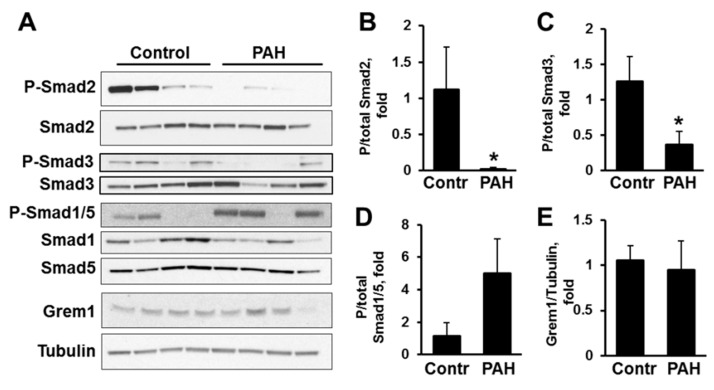
Increased growth and proliferation of distal pulmonary artery vascular smooth muscle cells (PAVSMC) is an important pathological component of pulmonary arterial hypertension (PAH). Transforming Growth Factor-β (TGF-β) superfamily plays a critical role in PAH, but relative impacts of self-secreted Activin A, Gremlin1, and TGF-β on PAH PAVSMC growth and proliferation are not studied. Here we report that hyper-proliferative human PAH PAVSMC have elevated secretion of TGF-β1 and, to a lesser extent, Activin A, but not Gremlin 1, and significantly reduced Ser-Smad2 and Ser-Smad3 phosphorylation compared to controls. Media, conditioned by PAH PAVSMC, markedly increased Ser-Smad2, Ser-Smad3, and Ser-Smad1/5 phosphorylation, up-regulated Akt, ERK1/2, and p38 MAPK, and induced significant proliferation of non-diseased PAVSMC. Inhibitory anti-Activin A antibody reduced PAH PAVSMC growth without affecting canonical (Smads) or non-canonical (Akt, ERK1/2, p38 MAPK) effectors. Inhibitory anti-TGF-β antibody significantly reduced P-Smad3, P-ERK1/2 and proliferation of PAH PAVSMC, while anti-Gremlin 1 had no anti-proliferative effect. PDGF-BB diminished inhibitory effects of anti-Activin A and anti-TGF-β antibodies. None of the antibodies affected growth and proliferation of non-diseased PAVSMC induced by PAH PAVSMC-secreted factors. Together, these data demonstrate that human PAH PAVSMC have secretory, proliferative phenotype that could be targeted by anti-Activin A and anti-TGF-β antibodies; potential cross-talk with PDGF-BB should be considered while developing therapeutic interventions.
远端肺动脉血管平滑肌细胞(PAVSMC)的生长和增殖增加是肺动脉高压(PAH)的重要病理成分。转化生长因子-β(TGF-β)超家族在 PAH 中发挥着关键作用,但自身分泌的激活素 A、Gremlin1 和 TGF-β 对 PAH PAVSMC 生长和增殖的相对影响尚未得到研究。在这里,我们报告说,高度增殖的人类 PAH PAVSMC 分泌的 TGF-β1 增加,在较小程度上分泌激活素 A,但不分泌 Gremlin 1,与对照相比,Ser-Smad2 和 Ser-Smad3 磷酸化显著降低。PAH PAVSMC 条件培养基显著增加 Ser-Smad2、Ser-Smad3 和 Ser-Smad1/5 磷酸化,上调 Akt、ERK1/2 和 p38 MAPK,并诱导非疾病 PAVSMC 的显著增殖。抑制性抗激活素 A 抗体可减少 PAH PAVSMC 的生长,而不影响经典(Smads)或非经典(Akt、ERK1/2、p38 MAPK)效应物。抑制性抗 TGF-β 抗体显著降低了 P-Smad3、P-ERK1/2 和 PAH PAVSMC 的增殖,而抗 Gremlin 1 则没有抗增殖作用。PDGF-BB 减弱了抗激活素 A 和抗 TGF-β 抗体的抑制作用。PAH PAVSMC 分泌的因子诱导的非疾病 PAVSMC 的生长和增殖不受任何一种抗体的影响。总之,这些数据表明,人类 PAH PAVSMC 具有分泌性增殖表型,可被抗激活素 A 和抗 TGF-β 抗体靶向;在开发治疗干预措施时,应考虑与 PDGF-BB 的潜在交叉对话。